Fetal Circulation
During intrauterine life, the mother ensures perfect nutrition for the fetus. The Placenta is the central organ of this nutrition. Its maternal part is formed by blood sinuses, and the fetal part by villi penetrating into the maternal part.
Fetal circulation before birth[edit | edit source]
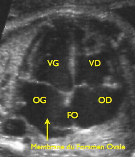
The blood circulation of the fetus' is modified so that there is an exchange of blood gases between the body of the fetus and the placenta, thus the placenta replaces the lungs of the fetus. Here, the blood is enriched with oxygen and nutrients and gets rid of waste products (CO2). Fetal hemoglobin here is saturated with oxygen to 60%, so it does not reach the saturation values of adult hemoglobin in the lungs (98%). And further, the blood bypasses the lungs, which are non-functional in the womb fetus.
The left and right sides of the fetal circulation communicate through two shunts that are right-left:
- foramen ovale - between the right and left heart atrium;
- ductus arteriosus - between the lung and the aorta (beyond the distance between the arteries supplying the LV and the head).
Blood from the fetal body is drained by paired aa. umbilicales (branches of the a. iliaca interna, after birth, their greater part disappears - the so-called chordae arteriarum umbilicalium seu ligg. umbilicilia medialia), these arteries enter the umbilical cordu (connection between the fetus and the placenta), after oxygenation and exchange of metabolites, the blood from the placenta is led to the body of the fetus again through the umbilical cord in the v. umbilicalis (it is unpaired - the umbilical cord therefore contains one umbilical vein and two umbilical arteries), v. umbilicalis takes place in the body of the fetus as the bottom edge of lig. teres hepatis from the navel to the liver (after childbirth, the v. umbilicalis changes to the lig. teres hepatis).
Some of the blood (almost half) goes to the liver. Because it is necessary to maintain a sufficient supply of oxygen and nutrients for the brain and other organs, a connection (short circuit) is created - ductus venosus Arantii (blood flow is regulated by a sphincter), which bypasses the liver and connects directly to the vena cava inferior (after birth, the ductus changes into lig. venosum Arantii) - thus oxygenated blood from the placenta flows into the heart inferior vena cava and its flow in the right atrium is directed against the foramen ovale to the left atrium by means of a valve - valva venae cavae inferioris Eustachi - the oxygenated blood then enters the arch of the aorta from the left heart and its branches mainly to the vessels of the head, neck and upper limbs.
Deoxygenated blood from the head, neck and upper limbs flows into the v. cava superior and on its way to the right atrium, where the blood flow is directed against the right atrioventricular opening via the transverse wall - torus intervenosus, continues from the right heart to the truncus pulmonalis and from it most of the blood reaches the aorta through the connector - ductus arteriosus Botalli''(this the clutch bypasses the lungs, which are collapsed and non-functional in the fetus - they are not sufficiently developed, due to the contracted pulmonary arteries there is high flow resistance andhypoxic vasoconstriction in them, only 10% of the fetal cardiac output flows through the lungs) , but only beyond the distance of the branches for the head, neck and upper limbs, the mixture of oxygenated and deoxygenated blood from the descending aorta then partly (35%) supplies the lower half of the trunk and lower limbs, partly (65%) flows to aa. umbilicales and through them again to the placenta.
- Summary
Thanks to the above-mentioned blood shunts, the best oxygenated blood flows from the "vena umbilicalis" through the inferior vena cava "(v. cava inferior)" to the heart and then through the "foramen ovale" to the left atrium and ventricle and subsequently into the aorta. Blood with high saturation thus reaches mainly the myocardium and the brain of the fetus.
Summary of the most important circulatory changes during childbirth[edit | edit source]
- Interruption of placental blood circulation;
- demise of the fetoplacental unit;
- the beginning of breathing through the lungs and associated changes in blood circulation.
Changes in fetal circulation after delivery[edit | edit source]
Foramen ovale[edit | edit source]
During labor, blood stops flowing through the placenta and the baby goes into hypoxia. The loss of oxygen and the increased partial pressure of CO2 in the blood will cause the irritation of the respiratory center in the CNS and cause the child's breathing movements. Negative interpleural pressure (−30 to −50 mmHg) also contributes to this. When the fetus passes through the birth canal, passive compression of the chest expels approximately 30 ml of fetal lung fluid from the lungs and trachea (the rest of the fetal lung fluid is resorbed). After birth, chest decompression occurs, which produces a small passive breath and is accompanied by an active glossopharyngeal effort. After the first breath, the lungs expand and a proportional amount of blood flows into them, which is already oxygenated here. Aeration of the lungs leads to the formation of surface tension, responsible for the retractive contractile force involved in passive expiration. The consequence of these changes is an increase in blood and lymphatic flow through the lungs. Stabilization of pulmonary ventilation induces stimuli to rebuild fetal blood circulation to neonatal circulation. bradykinin is released from the lungs, which is dependent on high blood oxygenation.
Bradykinin has a vasoconstrictive effect on the ductus arteriosus and umbilical vessels, and a vasodilating effect on the pulmonary vessels. Expanding the lungs reduces the resistance to blood flow to the lungs to less than 20% of the original, which increases blood flow through the arteriae pulmonales. This increases the amount of blood that returns to the left atrium. Pressure rises in the left atrium, causing the foramen ovale to close. The Foramen ovale disappears by the proliferation of endothelial and fibrous tissue by the third month, and in the adult heart it remains as the fossa ovalis'.
Ductus venosus Arantii – lig. venosum[edit | edit source]
The ductus venosus sphincter closes and the blood only penetrates into the hepatic sinusoids and via the venae hepaticae into the vena cava inferior. Later, it obliterates the ligament in the ligamentum venosum.
Ductus arteriosus Botalli – lig. arteriosum[edit | edit source]
The Ductus arteriosus already contracts during childbirth and with the first breaths it completely closes thanks to the influence of bradykinin. Anatomical closure occurs by the twelfth week, a part of the ductus is obliterated in the ligamentum arteriosum. By closing the foramen ovale and the ductus arteriosus, the blood circulation is definitively divided into pulmonary and systemic.
Umbilical vein[edit | edit source]
The ligamentum teres hepatis arises from the vena umbilicalis after birth
Umbilical arteries[edit | edit source]
The ``arteriae umbilicales give rise to the ligamenta umbilicalia medialia and the arteriae vesicales superiores, which supply the bladder.
Defective development of fetal circulation after birth[edit | edit source]
Foramen ovale patens[edit | edit source]
It occurs when the foramen ovale is not closed due to defective development of the septum primum and/or septum secundum. It is present in about 25% of people and usually has no effect on hemodynamics. However, in combination with another heart disorder, it can cause cyanosis of the skin and mucous membranes due to insufficient oxygenation.
Ductus arteriosus patens[edit | edit source]
Failure to close the ductus arteriosus or Botall's duct is the second most common congenital heart defect. It occurs 2–3 times more often in women. The first symptoms include increased respiratory effort and tachycardia. Ductus arteriosus patens can be idiopathic (without an induced cause) or affected by various factors such as: premature birth, congenital rubella syndrome, fetal alcohol syndrome or some chromosomal abnormalities (Down syndrome). Left untreated, it can lead to heart failure. It is treated with surgical ligature or with indomethacin without surgery.
Conclusion[edit | edit source]
Important changes occur during childbirth:
- cutting the umbilical cord definitively ends the function of the placenta;
- the beginning of breathing with lung expansion leads to a decrease in pulmonary vascular resistance;
- the increase in pulmonary blood flow results in an increase in pressure in the left atrium, which acts on the valve in the foramen ovale and this shunt closes;
- the flow in the right atrium is reduced and thus the "vena umbilicalis" is closed;
- left ventricular output resistance increases and the arteriae umbilicales close;
- the ratio of the thickness of the walls of the right and left ventricles changes significantly;
- the connection of the small and large circulation is parallel in the fetus, after birth it changes to serial.
Links[edit | edit source]
Related Pages[edit | edit source]
Source[edit | edit source]
- PASTOR, Jan. Langenbeck's medical web page [online]. [feeling. 04/09/2009]. < https://langenbeck.webs.com/ >.
References[edit | edit source]
- GANONG, William F. Přehled lékařské fysiologie. 1. edition. H & H, 1995. 681 pp. ISBN 80-85787-36-9.
- TROJAN, Stanislav. Lékařská fyziologie. 4., přeprac. a uprav edition. Grada Publishing, a. s, 2003. 772 pp. ISBN 80-247-0512-5.
- – GRIM, Miloš. Anatomie. 2. upr. a dopl edition. Praha : Grada Publishing, 2004. 673 pp. ISBN 80-247-1132-X.
- POCOCK, G – RICHARDS, C. D. Human Physiology : The Basis of Medicine. 1. edition. Oxford. 1999. ISBN 0192629522.
- SILBERNAGL, S – DESPOPOULOS, A. Atlas fyziologie člověka. - edition. Praha. 1993. ISBN 808562379X.
- wikipedia.com
- http://mcb.berkeley.edu/courses/mcb135e/fetal.html
- https://www.cayuga-cc.edu/blogs/
- https://medlineplus.gov/ency/article/001113.htm
- https://emedicine.medscape.com/article/891096-overview
- http://www.embryology.ch/anglais/pcardio/umstellung01.html
- https://brooksidepress.org/Products/Obstetric_and_Newborn_Care_1/lesson_2_Section_1D.htm


