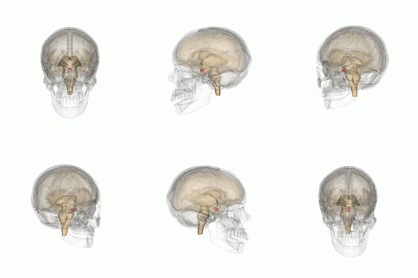
Disease of the adenohypophysis
The adenohypophysis is part of the hypothalamic-pituitary system, which is involved in the control of endocrine glands, growth and metabolism, as well as maintaining water and electrolyte balance. Disease of the pituitary gland is manifested both by symptoms of endocrine gland dysfunction and local symptoms related to the location of the adenohypophysis. As with other endocrine organs, there are either manifestations of hypofunction or excessive hormone secretion - hyperfunction.
Hypopituitarism[edit | edit source]
It is a hypofunction in which there is insufficient production of one or more hormones of the adenohypophysis. Panhypopituitarism means insufficient secretion of all hormones of the adenohypophysis.
Epidemiology[edit | edit source]
This is a rare disease.
Etiopathogenesis[edit | edit source]
Due to the functional connection with the hypothalamus, a disease of the hypothalamus or hypothalamo-pituitary stalk can manifest itself as hypopituitarism. The adenohypophysis is most often affected by pressure in the region of the ``Turkish saddle (pituitary adenoma, craniopharyngeoma, cysts, meningiomas, gliomas,...). Idiopathic hypopituitarism occurs more rarely. It can also be the result of radiation, trauma, bleeding, inflammation and others.
Clinical picture[edit | edit source]
The clinical picture depends on the amount of lost hormones and the depth of their deficit. If there is a gradual loss of hormone secretion, the order is usually as follows: LH/FSH → GH → TSH → ACTH. [1]
- Gonadotropin deficiency (LH and FSH) manifests as hypogonadotropic hypogonadism.
- GH (growth hormone) deficiency': growth retardation or arrest in childhood. In adulthood, an increase in fat mass, a decrease in physical activity, including cardiac performance, fatigue, a general deterioration of cognitive functions and a sense of health. Bone metabolism and the spectrum of lipoproteins deteriorate.
- TSH deficiency: central hypothyroidism.
- ACTH deficiency manifests itself as central hypocorticalism (secondary adrenocortical insufficiency). Deficiency of glucocorticoids dominates, secretion of mineralocorticoids tends to be sufficient.
- Prolactin deficiency is usually not manifested, because patients with its deficiency can no longer get pregnant due to the lack of gonadotropins.
Diagnostics[edit | edit source]
It includes a hormonal examination, which is different for each hormone, as well as a morphological imaging of the adenohypophysis (MRI) and a perimeter examination.
- GH Deficiency': we demonstrate using stimulation tests (insulin,...) when we observe an increase in GH secretion. We also determine IGF-I concentrations.
- Gonadotropin deficiency: we demonstrate using reduced levels of testosterone and estradiol with simultaneous unincreased levels of gonadotropins.
- TSH deficiency: we demonstrate a decrease in free T4 and at the same time an unincreased TSH concentration.
- ACTH deficiency: we demonstrate a reduced cortisol level and its insufficient increase during stress tests (insulin,...) and at the same time plasma ACTH levels are not increased.
- Prolactin deficiency: assessed by basal serum concentrations two hours after waking up.
Therapy[edit | edit source]
We treat hypopituitarism by replacing individual hormones of the peripheral glands (i.e. cortisol, thyroid hormones, estradiol, testosterone). The exception is hypogonadism, where we try to restore fertility, we have to administer gonadotropins. Growth hormone deficiency is also addressed by administration of human recombinant GH. GH is administered subcutaneously once a day, and treatment in adults is monitored by serum IGF-I concentrations. For children, we set the doses according to the calculated anthropometric parameters.
Prognosis[edit | edit source]
If the adenohypophysis is not damaged by a malignant tumor, the patient's prognosis is good.
Pituitary tumors[edit | edit source]
Benign adenomas are most often found in the pituitary gland. More rarely, craniopharyngioma, chordoma, meningioma, glioma and others can appear here. Pituitary adenomas are divided both according to their size and according to hormonal production.
Depending on the size, we differentiate:
- microadenomas – are less than <1 cm in greatest dimension
- macroadenomas – they are more than 1 cm
'According to hormonal production, we distinguish:
- prolactinomas - with overproduction of prolactin - 'the most common adenoma of the pituitary gland
- adenomas 'clinically hormonally afunctional - clinically afunctional, but hormonal production is often demonstrated immunohistochemically
- somatotropinomas – with overproduction of GH
- corticotropinomas – overproduction of ACTH
- gonadotropinomas – overproduction of LH or FSH
- thyrotropinomas – overproduction of TSH
Clinical signs[edit | edit source]
Clinical symptoms are divided into endocrinological symptoms and symptoms resulting from the oppression of surrounding structures.
- Endocrinological symptoms: result both from the overproduction of individual hormones and from the suppression of healthy pituitary tissue with a deficiency of one or more pituitary hormones.
- 'Symptoms from oppression of surrounding structures: oppression of the optical tract manifests itself as bitemporal hemianopsia, scotomas and even blindness. "Pressure on the hypothalamus" can lead to appetite disorders, thermoregulation disorders, diabetes insipidus and more. Expansion into the "sinus cavernosus" region results in diplopia, ptosis of the eyelid, ophthalmoplegia, disorders of facial sensitivity (n. III, IV, VI, V1 and V2 sub>). Pressure on the frontal and temporal lobes manifests itself as olfactory hallucinations and personality disorders.
Diagnostics[edit | edit source]
We determine the secretion of individual pituitary hormones in order to reveal their overproduction or, on the contrary, a decrease in their secretion. Morphologically, we examine the pituitary gland using MR. We complete an ophthalmological examination to determine the oppression of the optic tract.
Therapy[edit | edit source]
Therapy varies according to the size and hormone production of the adenoma. For prolactinomas, drug treatment is the first choice.'''''''''''''''''''''''''''''''''''''''''''''For other endocrine-active adenomas is neurosurgical removal of the adenoma. As for an afunctional microadenoma, it can only be observed. Afunctional macroadenomas are treated neurosurgically. In addition to the surgical solution, today, adenomas up to 3 cm can be successfully treated with radiation using the "Leksell gamma knife" (however, the distance from the optic tract depends).
Prolactinoma[edit | edit source]
Prolactinoma is the most common endocrine-active adenoma with an incidence of 30 cases per million inhabitants per year.
Etiopathogenesis[edit | edit source]
The etiopathogenesis is unknown, but it may occur within the MEN-I syndrome.
Clinical picture[edit | edit source]
In women of reproductive age, prolactinoma manifests itself in menstrual cycle disorders and infertility, and galactorrhea is common. In men, the symptoms are not very noticeable - a decrease in libido, erectile dysfunction, gynecomastia and, rarely, galactorrhea are common.
Differential diagnosis[edit | edit source]
It includes the differential diagnosis of hyperprolactinemia, which is quite broad. Hyperprolactinemia can arise from physiological causes (physical exertion, pregnancy, postpartum, idiopathic), it can also be caused by some drugs (especially psychotropic drugs), but it can also be part of multiple sclerosis, renal insufficiency, lupus, etc. A specific entity is the so-called pseudoprolactinoma , which is an afunctional pituitary adenoma that causes hyperprolactinemia by suppressing the pituitary stalk (thereby preventing the inhibitory effect of dopamine on secretion).
Therapy[edit | edit source]
The treatment of choice is drug therapy with dopamineu agonists, eg cabergoline or bromocriptine. Dopamine inhibits the secretion of prolactin and in up to 80% of patients leads to the normalization of prolactinemia and the reduction of the size of the adenoma.
![]() If an intense headache and visual disturbance appear at the beginning of the treatment, these are signs of bleeding into the tumor and a rapid neurosurgical solution is necessary!
If an intense headache and visual disturbance appear at the beginning of the treatment, these are signs of bleeding into the tumor and a rapid neurosurgical solution is necessary!
Prognosis[edit | edit source]
The prognosis is usually favorable. The treatment of oppressive macroprolactinomas, which are difficult to remove surgically and relatively resistant to radiation therapy, tends to be complicated.
Somatotropin[edit | edit source]
Somatotropinoma causing long-term and excessive secretion of GH leads to the development of acromegaly in adult patients and gigantism in children. A combination known as gigantoacromegaly can also occur.
Epidemiology[edit | edit source]
Acromegaly is a rare disease with an incidence of 4 cases per million population per year.
Etiopathogenesis[edit | edit source]
As already stated, the cause is a pituitary adenoma with overproduction of GH. It can also be adenomas with combined secretion of GH and prolactin. Acromegaly can be part of the MEN-I syndrome.
Clinical picture[edit | edit source]
In gigantism, excessive axial growth occurs because the growth fissures are not yet closed. Hypogonadism is also common. Acromegaly is characterized by ``enlargement of the acral body parts (ears, nose, lips, lower jaw, supraorbital arches) and a characteristic involvement of the fingers, where the proliferation of soft tissues creates the impression of blunt, peg-like fingers. It also includes enlargement of organs - 'organomegaly. Noticeable is macroglossia, which manifests itself in slurred speech. Fluid retention leads to swelling, especially of the fingers and feet. Hypertrophy also affects the sweat glands. The ``joint damage causing premature arthrosis is prominent. Patients' survival is negatively affected by the involvement of the cardiovascular apparatus. It is complex and includes arterial hypertension, accelerated atherosclerosis and acromegalic cardiomyopathy (hypertrophy, fibrosis, systolic and diastolic dysfunction). The resulting insulin resistance leads to the development of diabetes mellitus. Cephalgias are typical.
Diagnostics[edit | edit source]
Due to the episodic secretion of GH, we take 3 samples at hourly intervals to reliably demonstrate increased secretion. We also set endingsinjections of IGF-I. If we demonstrate increased GH secretion, MRI of the pituitary gland follows to demonstrate adenoma.
Therapy[edit | edit source]
The first choice is neurosurgical removal of the adenoma. If this is not possible or the removal is incomplete, we can use dopamine agonists to suppress GH production. However, treatment with superactive somatostatin analogues, e.g. lanreotide, is significantly more effective. If even this treatment does not sufficiently suppress GH production, we proceed to the use of growth hormone antagonists (pegvisomant). We also pay attention to the treatment of associated diseases.
Prognosis[edit | edit source]
With early treatment, the prognosis is favorable. If complications have already developed, patients have significantly increased morbidity and mortality.
Corticotropin[edit | edit source]
An ACTH-producing adenoma leads to the development of Cushing's disease.
Thyrotropin[edit | edit source]
An adenoma with overproduction of TSH leads to the development of hyperthyroidism with or without concomitant goiter. Diagnostics will demonstrate elevated levels of free T4 and TSH. Treatment is primarily neurosurgical, possibly radiological. In case of failure of the operation, it is also possible to use superactive analogues of somatostatin.
Gonadotropin[edit | edit source]
Adenomas with overproduction of LH and FSH usually have the clinical picture of non-functional adenomas and symptoms of oppression are manifested. Higher FSH levels and varying degrees of panhypopituitarism can be demonstrated in the laboratory. Treatment is again surgical with postoperative radiotherapy LGN.
Links[edit | edit source]
Related Articles[edit | edit source]
- Hypothalamic-pituitary system
- Diseases of the hypothalamic-pituitary system
- Pituitary function test
- Pituitary adenoma
References [ edit | edit source ][edit | edit source]
- ČESKA, Richard, et al. Internal 1st edition. Prague: Triton, 2010. 855 pp. ISBN 978-80-7387-423-0 .
- KLENER, Pavel, et al. Internal Medicine. 4th edition. Prague: Galén: Karolinum, 2011. 1174 pp. ISBN 978-80-7262-705-9 .
- LONGO, Dan L., et al. Harrison's principles of internal medicine. 18th edition. New York: McGraw-Hill, Medical Publishing Division, 2012. ISBN 978-0-07-174889-6 .
References [ edit | edit source ][edit | edit source]
- ↑ BUREŠ, Jan, et al. Internal Medicine 2. 2nd ed. Prague: Galén, 2014. p. 799. ISBN 978-80-7492-145-2 .
Category :Internal Medicine Endocrinology Pathology
- ↑
„ {{{1}}} “

