Determination of the number of cells, their viability and metabolic activity
Many tissue culture procedures require accurate determination of the number of cell in the sample. In practice, two methods of direct cell count are commonly used: manual counting in a calibrated chamber under a light microscope and semi-automatic counting using a particle flow counter.
Counting cells in a cell[edit | edit source]
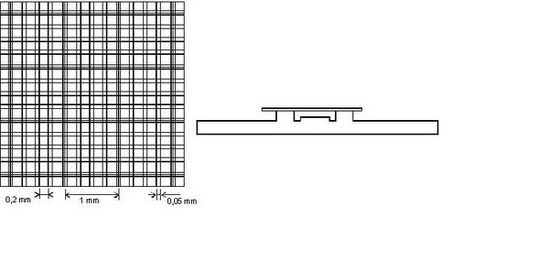
Counting cells in a cell is the simplest and often the most accurate method. We usually use the Bürker chamber. Two fields exactly 0.1 mm deep are ground on one microscope slide, on the bottom of which there is a net pattern (the distances of the individual lines are shown in the picture).
If it is necessary to know the number of live and dead cells in the sample, a dye that does not pass through the intact cell membrane or is actively transported out of the intracellular space is added to the cell suspension before counting. Trypan blue solution is most often used. Living cells remain colorless, while dead cells, where the integrity of the cell's membrane and its transport mechanisms are compromised, quickly turn blue.
Using a flow computer[edit | edit source]
When using a flow computer of particles, the device can be supplemented with other sensors that will more closely determine the character of the cells. Most often, the electrical properties of the particles are measured or their interaction with the laser beam is monitored. These techniques are referred to as flow cytometry and are routinely used in clinical biochemistry and hematology, for example to determine blood count, bone marrow examination or urinary sediment.
Other methods of determining the number of cells are also used:
- densitometry of stained cell cultures (dyes used in histology, methylene blue, Janus green);
- determination of DNA in the sample;
- determination of total protein in the sample.
- determination of the activity of some enzymes (hexose aminidase).
A special position among these techniques is occupied by MTT test. MTT (3-[4,5-dimethylthiazol-2-yl]-2,5-diphenyltetrazolium bromide) is reduced to colored formazan by mitochondrial dehydrogenases. The rate of formazan formation corresponds to the activity of the respiratory chain, and thus reflects the metabolic activity of the cell.


