Congenital heart defects/Repetitorium
Congenital heart defects[edit | edit source]
Embryonic septation of the heart
Classification[edit | edit source]
- Without shunt: coarctation of the aorta, valvular defects (pulmonary stenosis, aortic stenosis)
- With a shunt:
- with possible cyanosis (left-right shunt: ostium secundum atrial septal defect - ASD, ventricular septal defect - VSD, atrioventricular septal defect - AVSD, patent ductus arteriosus - PDA)
- cyanotic (right-left shunt: tetralogy of Fallot - TOF, transposition of the great arteries - TGA, defects with a common chamber - single ventricle type defect - SV, hypoplastic left heart syndrome - HLHS)
Factors determining the magnitude and direction of shunt flow[edit | edit source]
- pressure gradient
- shunt location
Cyanosis[edit | edit source]
Cyanosis occurs when the value of reduced hemoglobin rises above 50g/l. This happens in complex defects with a shunt, when oxygenated and non-oxygenated blood mixes. In these situations, the amount of oxygenated blood in the "mixture" depends on the amount of pulmonary flow. In cases where the lesion is not surgically corrected soon, compensatory polyglobulia occurs and the degree of cyanosis may not correspond to the severity of the defect.
Consequences of a left-right shunt[edit | edit source]
- reaction of the pulmonary canal
- shunt reversal: Eisenmenger syndrome
Examples of congenital heart defects[edit | edit source]
Coarctation of the aorta[edit | edit source]
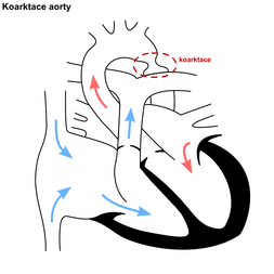
The arch of the aorta is narrowed, most often behind the distance of the left subclavian artery, but sometimes it can also be between the distances of the large vessels, which changes the value of the systolic pressure and the strength of pulsation in the upper and lower extremities, respectively in the right and left HR. This condition puts pressure on the left heart and at the same time develops a condition where the upper half of the body is perfused normally, while the lower half suffers from hypoperfusion.
Tetralogy of Fallot[edit | edit source]
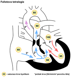
Tetralogy of Fallot is a combination of four defects: right ventricular outflow tract stenosis, ventricular septal defect, aorta overlying the defect, and right ventricular hypertrophy. Due to the increased pressure in the right ventricle, a right-left shunt occurs at the level of the ventricular septal defect, which is manifested by cyanosis. Tetralogy of Fallot is characterized by so-called hypoxic attacks, when, due to stress, the outflow tract of the right ventricle contracts even more, pulmonary flow decreases and cyanosis becomes more pronounced.
Open Botallo's duct[edit | edit source]
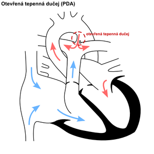
Botallo's duct is a communication between the aorta and the lung, which prenatally allows blood to flow from the high-pressure pulmonary basin to the systemic one. After birth, blood saturation in the duct increases, which leads to its spontaneous closure. If this does not happen, due to the rapid decrease in pressure in the pulmonary artery, the shunt turns and blood flows from the aorta back into the pulmonary artery, with all the consequences for the pulmonary circulation.
Septal defects[edit | edit source]
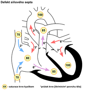
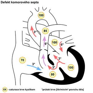
Septal defects occur at the level of the atrial septum (ostium primum defect), atrioventricular septum (AVSD, ostium secundum defect) and the membranous or muscular part of the ventricular septum. Blood flows through the shunt in the direction of the pressure gradient at the level of the shunt.
Links[edit | edit source]
Related articles[edit | edit source]
Source[edit | edit source]
- VÍZEK, Martin. Repetitorium [online]. [cit. 2011-11-12]. <https://web.archive.org/web/20130512032641/http://pf.lf2.cuni.cz/vyuka/repetitorium.html>.
References[edit | edit source]
Literature[edit | edit source]
- OŠŤÁDAL, Bohuslav – VÍZEK, Martin, et al. Patologická fyziologie srdce a cév. 1. edition. Praha : Karolinum, 2003. ISBN 80-246-0597-X.

