Burkitt lymphoma

Burkitt lymphoma, in the French literature also le lymphoma non-Foltys, is a highly malignant tumor classified as non-Hodgkin's lymphomas. Its structure is formed by mature B-cells. It typically occurs in the lower jaw area. It mainly affects children and young adults.
Pathogenesis[edit | edit source]
Tumor cells have the characteristics of B-lymphocyte. From a genetic point of view, they are characterized by translocation of the c-myc protoncogene from chromosome 8 to 2, 14 or 22 into the region encoding the immunoglobulin chains. In 80% of cases, it is a balanced translocation from chromosome 8 to 14 [1]. By changing the position of c-myc its unregulated expression occurs. Because the gene encodes a transcription factor that stimulates cell division genes, there is an uncontrolled proliferation of cells .
Forms[edit | edit source]
Lymphoma occurs in three different forms:
- associated with immunodeficiency (often in patients with AIDS, patients have infiltrated bone marrow and nodules),
- sporadic (most often manifested as an intra-abdominal tumor mass in the ileocecal region, the kidneys and ovaries may be affected, adults tend to have infiltrations in the nodes),
- endemic.
The endemic form occurs in equatorial Africa and New Guinea [2]. In these areas, it is one of the most common cancers in children [1]. In 50% of cases, it affects the bones of the face, it can occur in the gastrointestinal tract (small and large intestine), paired abdominal organs (ovaries) and in the breast, often bilaterally. In the development of an endemic form, a connection with infection EBV is evident.[3]. Patients often have several-fold higher levels of antibodies to EA (early nonstructural antigens) and VCA, with viral DNA and EBNA present in tumor cells. EBV-associated lymphoma develops 7–31 months [4] after the acute phase infectious mononucleosis. Levels of anti-EA antibodies rise before lymphoma develops and signal activation of latent infection.
- Soubor:Burkitt's lymphoma.jpg
Burkitt's lymphoma of the jaw
- Soubor:Malignant Burkitt’s lymphoma, a non-Hodgkin’s lymphoma (NHL) 064 lores.jpg
Burkitt lymphoma
Microscopic image[edit | edit source]
Histologically the tumor is uniform. A reduced amount of cytoplasm, which is basophilic, is enclosed in cells with blurred contours of the cytoplasmic membrane. Dark tumor cells form the background for bright macrophages, giving the impression of a "starry sky".
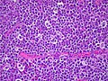
Burkitt lymphoma, dyeing hematoxylin-eosin
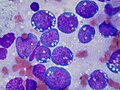
dyeing Wright


Therapy[edit | edit source]
The disease is well treatable, with a good prognosis. The outcome of the treatment depends on staging.