Biophysics of hearing
Hearing is the ability of animals to perceive sound using a specialized organ - the ear. We distinguish two types of sound conduction: bone conduction and air conduction.
Air conduction is the conduction of sound through the ear canal – ear drum – auditory ossicles – oval window. In addition, it is possible to oscillate the fluids in the inner ear by direct transmission of vibrations of the skull bones - in this case we are talking about bone conduction. The hearing threshold for bone conduction in a healthy person is about 40 dB higher than the threshold for air conduction, so bone conduction is mainly used where air conduction is impaired. A healthy person uses bone conduction when perceiving his own voice or very strong sounds.
The human ear is able to perceive sounds with a frequency of 16 Hz–20 kHz , a level of 0 dB (hearing threshold) to 130 dB (pain threshold).
Brief anatomy of the auditory system[edit | edit source]
For a correct understanding of the hearing process and for a simpler description of the individual phases of sound conduction by the ear, it is necessary to at least lightly outline the anatomical structure.
The human ear consists of three parts:
- External ear (auris externa)
- Middle ear (auris media)
- Inner ear (auris interna)
External ear[edit | edit source]
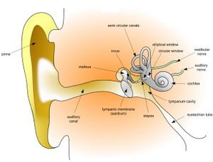
The external ear consists of the pinna (auricula), the external ear canal (meatus acusticus externus) and the ear drum (membrana tympani).
The auditory pinna is rudimentary in humans and its function is minimal. Unlike other mammals, the muscles of the human auricle are without functional significance. Their innervation is from n. VII.
The external ear canal is a partly cartilaginous and partly bony tube, beginning at the porus acusticus externus and ending at the eardrum. Its base is the os tympanicum. The narrowest point (isthmus) of the external ear canal is at the interface between the cartilaginous and bony sections and has a diameter of 6–8 mm. The total length of the external ear canal is 24–35 mm.
The tympanic membrane separates the external ear canal from the middle ear cavity. It is a thin oval membrane (9 x 10 mm) with a thickness of 0.1 mm. The auditory ossicle, the malleus, connects to the eardrum in the middle ear.
Middle ear[edit | edit source]
The middle ear is located in the middle ear cavity (cavitas tympanica). It contains 3 auditory ossicles: malleus, incus, stapes.
The auditory ossicles are interconnected. The malleus is attached to the eardrum, followed by the anvil, on which the stirrup fits, which is connected to the membrane of the fenestra vestibuli (fenestra ovalis). The middle ear is connected to the nasopharynx by the Eustachian tube (tuba auditory). Auditory muscles also play an important role in the middle ear: the tensor tympani muscle (innervation of the V and VII nerves ), m. stapedius (innervation of n. VII ).
Inner ear[edit | edit source]
The inner ear includes a bony labyrinth (labyrinthus osseus) and within it a membranous labyrinth (labyrinthus membranaceus), which contains endolymph. The labyrinth has an equilibrium part, consisting of the vestibule and three semicircular canals, and an auditory part, which is represented by a bony and membranous cochlea (cochlea) with a receptive auditory organ of Corti, located on the basilar membrane (length about 3 cm). The auditory pathway runs from the ganglion cochleare to the upper part of the temporal lobe (the convolutions of Heschl).
Structue of the organ of Corti[edit | edit source]
The organ of Corti (organum spirale) is a complex system of supporting and sensory cells. The basis of the organ is two rows of supporting cylindrical pillar cells (cells of Corti), which together form the tunnel of Corti.
Sensory (hair) cells are located on either side of the pillar. Medially there is one row (inner hair cells), laterally there are three to four rows of hair cells (outer hair cells).
There are 1,500 inner hair cells. Their apical surface contains 50–60 stereocilia , which are in contact with the membrana tectoria. Outer hair cells are found in the number of 12–15,000, they are also equipped with stereocilia. In the basal part, they are in contact with afferent and efferent fibers.
Biophysics of hearing[edit | edit source]
The sound wave is directed through the pinna into the external auditory canal. The pinna, as already mentioned, is rudimentary, and its loss will not fundamentally affect hearing. The external auditory meatus carries the captured sounds to the eardrum, which impinges on it and vibrates it.
The deflections of the eardrum are very small (at a frequency of 1 kHz, about 10 −11 m). The area of the eardrum is about 55 mm2 and the area of the membrane window to which the vibrations are brought by means of the ossicles is only 3 mm2.
If we assume that the energy passing through both surfaces is the same, the acoustic pressure reaches the surface of the oval window many times greater (about 22x). This is necessary to overcome the acoustic resistance of the fluid in the cochlea. As auxiliary systems, the Eustachian tube and ear muscles are used in the middle ear, which equalize the pressure on the eardrum from the inside to prevent it from rupturing.
Oscillation of the oval window is caused by vibrations in the endolymph (incompressible fluid), which are further amplified by vibrations from the bone conduction, which reaches it through the bones of the skull . With its vibrations, the endolymph vibrates the membrana tectoria, which subsequently irritates the stereocilia of the inner hair cells. These release a small amount of mediator (probably glutamate) in the basal part of the cell, which creates a nerve signal.
The outer hair cells have an amplifier function. When they are irritated, there is an elongation and subsequent contraction of the cells, which increases the sensitivity of the inner hair cells. This mechanism allows very quiet sounds to be heard.
Links[edit | edit source]
External links[edit | edit source]
References[edit | edit source]
- NAVRÁTIL, Leoš – ROSINA, Jozef, et al. Medicínská biofyzika. 1 (dotisk 2013) edition. Grada Publishing, 2005. 524 pp. pp. 287-290. ISBN 978-80-247-1152-2.
- DRUGA, Rastislav – GRIM, Miloš. Anatomie periferního nervového systému, smyslových orgánů a kůže. 1. edition. Galén : Karolinum, c2013. pp. 135-153. ISBN 978-80-7262-970-1.
- ČIHÁK, Radomír. Anatomie 3. 2. edition. Grada Publishing, 2004. 692 pp. pp. 621-640. ISBN 978-80-247-1132-4.
- AMLER, Evžen. Acoustics [lecture for subject Biophysics, specialization General medicine (czech), 2. LF Charles University]. Prague. 2013.


