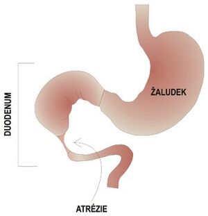
Atresia and stenoses of the small intestine
Duodenal atresia and stenosis [ edit | edit source ][edit | edit source]
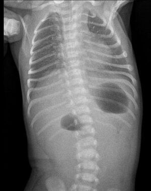
Duodenal atresia A typical finding on an X-ray image in the suspension of duodenal atresia - the so-called double bubble sign
The incidence of atresia and stenoses is 1:5000-10000. 30% of patients also have Down syndrome and more than 50% of patients have associated birth defects.
Clinical course [ edit | edit source ][edit | edit source]
Prenatally, polyhydramnios occurs due to interrupted circulation of amniotic fluid. The so-called image of two "bubbles" that are closed by the liquid occurs. Postnatally , with complete obstruction in the first 24 hours, the clinical picture of high ileus develops , which is manifested by violent vomiting with bile admixture. In 90% of cases, the obstruction is located below the papilla of Vater. In the remaining 10% of cases, the vomit does not contain bile. The doctor can observe a bulging epigastrium, sunken hypogastrium (boat-shaped belly), peristalsis is visible. Smolka is not leaving. In case of partial obstruction, the clinical manifestation is late.
Diagnostics [ edit | edit source ][edit | edit source]
Prenatal diagnosis is performed using ultrasound screening. Postnatally, a native X-ray of the abdomen is observed in the suspension, which is typically depicted as two bubbles or levels , this applies to the stomach and the enlarged duodenum. In most cases, there is an absence of gas in the distal part of the GIT . More than 20 ml of liquid is aspirated from the stomach using a nasogastric tube. The normal volume of fluid in the stomach is 5 ml. This is followed by insufflation of gas into the stomach and thanks to this the image of the two bubbles can be reproduced on the X-ray. Contrast X-ray of the upper part of the GIT is performed to clarify the diagnosis and at the same time rule out malrotation or volvulus.
Treatment [ edit | edit source ][edit | edit source]
The treatment is surgical, the patient undergoes a duodenoduodenal anastomosis.
Atresia and stenosis of the jejunum and ileum [ edit | edit source ][edit | edit source]
The incidence of atresia and stenoses of the jejunum and ileum is 1:1500.
Clinical course [ edit | edit source ][edit | edit source]
Polyhydramnios occurs prenatally . Postnatally, in the first 36 hours, the development of the clinical picture of a moderately high ileus occurs, which results in vomiting with bile. The abdomen is distended and there are also breathing difficulties - dyspnoea , due to the high state of the diaphragm. Smolka is not leaving. Dehydration with hypochloremia and weight loss develops.
Diagnostics [ edit | edit source ][edit | edit source]
Prenatal ultrasound shows dilatation of intestinal loops. Postnatally, a native X-ray of the abdomen in suspension is observed.
Treatment [ edit | edit source ][edit | edit source]
The treatment is surgical, the atretic or stenotic section of the intestine is removed and an end-to-end anastomosis is performed.
- Gallery of diagnostic X-ray images in patients with various obstructions of the small intestine
Links [ edit | edit source ][edit | edit source]
Related Articles [ edit | edit source ][edit | edit source]
- Congenital atresias and stenoses of the gastrointestinal tract
- Superior mesenteric artery syndrome
- Bowel malrotation and volvulus
- Meconium ileus
- Megacolon congenitum
References [ edit | edit source ][edit | edit source]
ŠNAJDAUF, Jiří and Richard ŠKÁBA. Pediatric surgery. 1st edition. Prague: Galén, 2005. ISBN 807262329X .
HOLCOMB III, George W., J. Patrick MUPRHY, and Daniel J. OSTLIE. Ashcraft's Pediatric Surgery. 6th edition. Elsevier, 2014. ISBN 145574333X .
MUNTAU, Ania Carolina. Pediatrics. 4th edition. Prague: Grada, 2009. p. 365. ISBN 978-80-247-2525-3 .


