Apoptosis and clinical consequences of disorders of its regulation
Apoptosis is a physiological process in which energy is consumed. It does not cause an inflammatory reaction (unlike necrosis). It is important for embryonic development, hormone-dependent cycles (endometrium, mammary gland, prostate) and cell renewal (intestinal epithelium, etc.). Disorders in the regulation of apoptosis can lead to tumours, hypotrophy of organs (e.g. kidney) or congenital malformations (e.g. syndactyly).
Stages of apoptosis[edit | edit source]
- Signal for apoptosis - the decision to die,
- execution of apoptosis,
- degradation of apoptotic bodies by phagocytosis.
Signal for apoptosis[edit | edit source]
- It can come by external or internal route,
- external pathway - lack of growth factors, cytotoxic cytokines (FasL, TNF), morphogens, glucocorticoids, etc.,
- internal pathway - signals come mainly from mitochondria, controlled by the Bcl-2 gene family (BAX and BCL-2 gene.
- Survival signals
- Growth factors, cytokines, hormones, viral protein p53.
- Signals for apoptosis
- Morphogens, viruses glucocorticoids, genotoxic influences, cytotoxic cytokines (TNF, FasL), T-lymphocytes and NK-cells (granzyma B, perforins).
- Genes - suppressors of apoptosis
- Bcl-2, bag, Rb.
- Genes - inducers of apoptosis
- bax, bak, bad, TP53.
Execution of apoptosis[edit | edit source]
- Apoptosis signals cascade ICE (caspase), which triggers the actual execution of apoptosis,
- chromatin condensation and its aggregation in the periphery of the nucleus, DNA cleavage between nucleosomes (fragments are 180 to 200 bp long),
- ER, increases, forms pockets and connects with the cytoplasmic membrane,
- vacuoles are formed and filaments are aggregated,
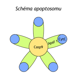
- mitochondria burst and release cytochrom C,
- In the end, apoptotic bodiesare formed.
Phagocytosis of apoptotic bodies[edit | edit source]
- Absorption by macrophages and surrounding cells.
[edit | edit source]
The Rb gene produces the protein p105, which is phosphorylated by cyclin/cdk complexes (cyclin D/cdk 4, 6 and cyclin E/ckd2) at least 10 sites at the G1 checkpoint, thereby altering its ability to associate with the E2F protein, which forms heterodimeric complexes with DP1. E2F and DP1 are transcription factors that induce the expression of genes necessary for the development of S-phase (e.g. DNA-polymerase, thymidine kinases, dihydrofolate reductase, c-myc, c-myb and cdc2). This means that the Rb gene is necessary for the development of the S-phase and suppresses apoptosis. Mutations in the Rb gene cause retinoblastoma, tumors of the bones, breasts, lungs, prostate or bladder.
The TP53 gene is a tumor suppressor gene whose key role is also to regulate the G1 checkpoint. The product of the TP53 gene, the p53 protein, is a transcription factor that activates gene expression for factors inhibiting cell proliferation. One of them is the p21 protein, which stops the development of the cell division cycle so that DNA repair can occur. If DNA damage is non-reparable, p53 is involved in apoptosis induction. TP53 mutations are present in 50% of tumors, congenital mutation of this gene leads to the development of Li Fraumeni syndrome.
Bcl-2 and bax are genes whose products form both homodimers and together heterodimers. Bcl-2 suppresses apoptosis, bax induces. Bcl-2-bax heterodimers do nothing. Depending on whether the amount of homodimers bax-bax or bcl2-bcl-2 predominates, apoptosis will occur or not.
The p35 protein is encoded by some viruses. It inhibits apoptosis so that the virus can continue to live and spread in the host cell. The p35 protein is cleaved by caspases instead of other substrates, thus preventing them from effectively inducing apoptosis.
Cells also need stimulation by survival factors (hormones, cytokines, growth faktors) to live. If they are not stimulated by them, this leads to apoptosis. These include, for example, PDGF, FGF, HGF, IGF, etc.
The Fas receptor is a transmembrane receptor that belongs to the TNF family. The transmission of the signal from it is through caspases. FasL is a transmembrane protein, but it can alternatively be cleaved from the membrane. FasL is expressed by T-lymphocytes, macrophages and NK-cells. Fas-induced apoptosis results from contact between the Fas receptor and FasL (ligand). This type of apoptosis is involved in the selection of T-lymphocytes in peripheral blood, the cytotoxic effect of T-cells and NK-cells, and the termination of the immune response (death of immune cell clones).
Caspases are proteases that are involved in various pathways of apoptosis induction. So far, 11 have been described. They are synthesized as precursors and can be activated e.g. via Fas receptors or TNF receptors, etc. After activation, cascade activation of caspases occurs (similar to e.g. hemocoagulation reaction), which leads to the execution of apoptosis.
Links[edit | edit source]
Related Articles[edit | edit source]
Bibliography[edit | edit source]
- KAPRAS, Jan and Milada KOHOUTOVÁ. Chapters from Medical Genetics III.. 1st edition. Praha : Karolinum, 2009. vol. 1. ISBN 80-246-0001-3


