Aorta summary
The aorta is a vessel emerging from the left ventricle as an ascending aorta (aorta ascendens), then curves to the left as an aortic arch (arcus aortae) and continues caudally as a descending aorta (aorta descendens). It has two parts: the thoracic aorta and the abdominal aorta. Along the way, the aorta releases direct branches nourishing the surrounding organs.
Aorta thoracica[edit | edit source]
The thoracic aorta is the thoracic part of the descending aorta. It follows the arcus aortae at the height Th3 − Th4 and then runs first at the left side of the vertebrae, gradually gets in front of them and continues caudally. After passing through the aortic hiatus, the diaphragm at the Th12 level continues as the abdominal aorta. In front of the aorta lies together with the esophagus, which is slightly to the right, then cranially radix pulmonis sinistri and caudally heart. The thoracic ductus runs between the aorta and the esophagus. From the sides, the aorta is surrounded by the mediastinal pleura, through which the aorta is imprinted into the left lung. The posterior mediastinal pleura passes through the intercostal dorsal artery and sinus artery. It supplies the muscles of the posterior three quarters of 3. − 11. intercostal space, anterior part of abdominal muscles, part of diaphragm, skin on sides and back of chest, lungs, mediastinal organs, spinal canal, spinal cord and spinal cord envelopes.
The thoracic aorta, like the abdominal aorta, has branches for the surrounding walls and organs.
Accordingly, the following are recognized:
- parietal (wall) branches;
- visceral (organ) branches.
The parietal branches of the thoracic aorta are paired. Wall branches include:
- superior phrenic artery
- the superior phrenic artery protrudes above the diaphragm aorticus of the diaphragm and supplies its adjacent section;
- posterior intercostal arteries.
- Nine pairs of posterior intercostal arteries for 3rd − 11. the intercostal space gradually emerges from the aorta from behind and passes through the posterior mediastinal pleura. They follow the veins and nerves along the spine to the intercostal space and then further between the intercostal muscles interni et intimi, in the sulcus costae they go along with the veins (cranially from the artery) and nerves (caudally from the artery). In the anterior section of the intercostal space, they interast with the intercostales anteriores of the internal thoracic artery. During its course, it deletes aa. intercostales posteriores the following branches:
- r. dorsalis;
- r. collateralis;
- r. cutaneus lateralis;
- rr. lateral mammals.
The visceral branches are unpaired and protrude from the front of the aorta. Is part of them:
- rr. bronchiales - are 2-3 arteries arising above the aorta at the level of the bifurcation of the trachea (Th4−5). As a rule, two go to the left and one to the right, joining the bronchi and branching into the lungs;
- rr. oesophage;
- rr. pericardium;
- rr. mediastinals.
Aorta abdominalis[edit | edit source]
The abdominal aorta forms an unpaired continuation of the thoracic aorta and transports oxygenated blood to all abdominal and pelvic organs, supplying the muscles of the back, abdominal walls and diaphragm, external genitalia and lower limbs. The abdominal aorta runs close to the spine along the left flank of the inferior vena cava from the diaphragm aorticus (Th12 level) to its bifurcation (L4), where it divides into the iliacae communes (a. Iliaca communis dextra et sinistra) , which then continue to pelvic area, where it further branches. The abdominal aorta has several groups of branches. The basic aspect of the division is the fact that some branches supply the organs, while others participate in the supply of the surrounding walls. Accordingly, we recognize:
- parietal (wall) branches;
- visceral (organ) branches.
The parietal branches of the abdominal aorta include aa. phrenicae inferiores extending just below the aortic hiatus and extending along the lower surface of the diaphragm, 4 pairs of aa. lumbales, a. sacralis mediana forming an unpaired continuation of the abdominal aorta and aa. common iliacae. Aa. Phrenicae inferiores are involved in the supply of the diaphragm and also contribute to the nutrition of the adrenal glands (aa. suprarenales superiores). The lumbar arteries are a continuation of the thoracic intercostal arteries (branches of the thoracic aorta) and supply the corresponding sections of the lumbar region and abdominal wall. Aa. iliacae completely supply the lower half of the body.
Visceral branches of the abdominal aorta can be further divided into:
- paired;
- unpaired.
Typical unpaired branches include craniocaudally:
Truncus coeliacus is a very short branch, which is only a few centimeters from its distance in the Th12 / L1 area divided into 3 main branches - a. splenica supplying the large gastric curvature , body and tail of the pancreas and spleen, a. gastrica sinistra running along the small gastric curvature and supplying the pars abdominalis of the esophagus and a. hepatica communis nourishing the area of the large curvature, duodenum, pancreatic head and liver with gallbladder.
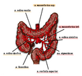
A. mesenterica superior (spaced about 2 cm caudally from the truncus coeliacus behind the head of the pancreas, level L1) is the main branch for the duodenum (aa. Pancreaticoduodenales inferiores), jejunum (aa. Jejunales), ileum(aa. Ileales), caecum (a. ileocolica), colon ascendens and colon transversum (a. colica dextra) up to the Cannon-Böhm point (about 2/3 colon transversum). Outside the [small|small intestine]], appendix and large intestine, the mesenteric artery it is also involved in the supply of the head of the pancreas, it can send branches also for the stomach and in up to 30% of cases even an additional hepatic artery.
A. mesenterica inferior (distance at the level of the upper part of the L3 vertebra) is followed by a supply to the. (anastomosis with a. colica media - anastomosis magna Haleri) and supplies blood for the rest of the colon transversum, for the colon descendens (a. colica sinistra), sigmoid (aa. sigmoideae) and rectum (a. rectalis superior), where it anastomoses with paired aa . rectales mediae (from the internal iliac artery), which allows partial (but not entirely sufficient) compensation in the event of obstruction of one of these arteries.
The paired branches of the abdominal aorta are:
- aa.suprarenales mediae supplying the right and left adrenal glands;
- aa.renales (a.renalis dx. et sin.) for both kidneys and as a lower branch for the adrenal glands (aa. suprarenales inferiores);
- aa.testiculares/ovaricae (a. testicularis / ovarica dx. et sin.) nourishing the gonads (testicles / ovaries).


