T cells
T-cells are white blood cells that are the part of the immune system. T-cells develop from the precursors in the thymus. These precursors migrate from the hematopoietic tissues (mainly from the bone marrow) during prenatal development.
In the thymus, the immature T-cells are selected. Reticular epithelial cells present antigens to the immature T-cells. The survival of the T-cell depends on their ability to distinguish HLA of the own body. Non-reactive and overly reactive cells are destroyed (about 95%). The T-cells that aren't destroyed enter the bloodstream. Blood transfers them to the secondary lymphoid organs, where they meet their specific antigens. This leads to their activation and subsequent inflammation development.
The Specificity of the T-cells is determined by the development of their TcRs (T-cell receptors). However, these alone are not enough to activate the lymphocyte (signal transmission), the membrane complex of TcR and CD-3 is necessary .
After the immune response has ended, the lymphocytes circulate between the blood and the secondary lymphatic tissues and wait in the resting form for another encounter with the antigen.
According to the function of individual T-lymphocytes and according to the CD markings that occur on their surface, we distinguish the following basic groups of T-lymphocytes:
T-helper cells[edit | edit source]
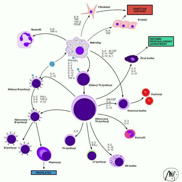
CD4+ cells produce many different cytokines. They react to the antigen-MHC class II complex presented by the APCs. They initiate a specific immune response. Based on the produced cytokines T-helper cells are divided into:
- T1: activates the cytotoxic and cell immune reaction (macrophages, TC).
- T2: activates the antibody reaction (B-cells, antibody production).
T-helper cells are the target of HIV.
Cytotoxic T-cells[edit | edit source]
CD8+, are able to cause apoptosis of the cell, or can destroy the cell directly by cytotoxic mechanisms. They react to the MHC class I. They control the condition of the cells in the body (anti-tumor immunity).
Suppressor T-cells[edit | edit source]
A non-uniform group of the T-cells, during the inflammation most likely develop from the T-helper cells, that's why they're also sometimes called as TH3(r) subset cells. They don't have the specific CD marker, some are CD-4+ and CD-8+, others carry either one of these markers or none of them. Their main task is to moderate and suppress the immune response. It is implemented by interleukins IL-10 and partially IL-4. Tissue reparation and fibroblast stimulation is accelerated by TGF-β (transforming growth factor).
Natural killer T-cells, NKT[edit | edit source]
The main components of the cytotoxic cell immunity. They are able to destroy even without prior encounter with the antigen (this applies to newborns). Although they're lymphocytes, they belong to the innate immune system. They do not carry the CD-3 character. Morphologically they're large lymphocytes (12–14 μm).
Related Articles [ modify | edit source ][edit | edit source]
Bibliography[edit | edit source]
- KONRÁDOVÁ, Václava – UHLÍK, Jiří – VAJNER, Luděk. Funkční histologie. 2. edition. Jinočany : H & H, 2000. 291 pp. ISBN 80-86022-80-3.
- ŠTERZL, Ivan. Základy imunologie pro zubní a všeobecné lékaře. 1. edition. Praha : Karolinum, 2005. 207 pp. ISBN 978-80-246-0972-0.

