Peripheral vein cannulation

Motto: If I'm trying to avoid something, I'm looking for a reason. If I'm trying to figure something out, I'm looking for a way.
Indications[edit | edit source]
- Securing potentially at-risk patients,
- provision for diagnostic purposes (eg for iv application of contrast),
- infusion therapy,
- blood transfusion ,
- parenteral medication administration,
- parenteral nutrition ,
- providing access for the introduction of a central venous catheter (CVC) from the periphery .
Contraindications[edit | edit source]
Contraindications include infection or injury at the puncture site or the presence of an arteriovenous shunt on the limb. A relative contraindication may be severe damage to the proximally placed veins, ignorance of the technique.
Venous access[edit | edit source]
For cannulation, we usually use short cannulas with a diameter of 14–26 G , which we insert into the superficial veins on the dorsum of the hand, in the cubital fossa, and less commonly on the wrist, forearm, dorsum of the foot. In children up to the age of about 2, surface veins on the calva that do not have valves can also be cannulated → the cannula can be inserted in any direction (but it is still better centripetally and in respect to the laws of gravity). We use the veins on the volar side of the wrist only exceptionally - puncture or other manipulations in this area are really painful procedures. In a situation of great peril we can cannulate v. femoralis , v. jugularis externa or v. axillaris.
Overview of venous access points[edit | edit source]
- Head + neck
- v. supraorbitalis, v. temporalis, v. auricularis posterior, v. occipitalis, v. jugularis interna, v. jugularis externa, v. subclavia.
- Upper limb
- v. axillaris, v. cephalica, v. basilica, plexus venosus dorsalis manus.
- Lower limb
- v. femoralis, v. saphena magna, v. saphena parva, plexus venosus dorsalis pedis.
Technique[edit | edit source]
It is important to be calm and patient during the procedure. Puncture success is significantly affected by detection of the vein and preparation for the puncture. The conditions for a successful try can be improved by warming the limb (causes vasodilation), "knocking" (toning of the venous wall) or pumping of blood into the venous system when the limb is constricted (increasing the filling of the venous system). The relevant limb / head is fixed by an assisting nurse, who ties up the limb above the injection site with a rubber "tourniquet" to increase the venous filling. After filling the venous system, we insert the cannula inserted on the needle mandrel so that the needle hole points in the direction of blood flow = centripetally, the needle should form an angle of 10-30 ° with the body surface (more tangentially, i.e. at a lower angle for a new-born on the volar side of the wrist or forearm). The technique of inserting the cannula itself also depends on the size of the cannula and the type according to the manufacturer - e.g. For cannulas of type Braun 22 G right after blood appears in cannula, we pull the mandrel and continue with insertion of only our own cannula, it is better to insert Braun 24G without pulling on the mandrel, i.e. to "pull" the cannula along the mandrel into a vein or to continue the final insertion after the initial light insertion with a small amount of solution. For cannulas with a larger needle overhang above the end of the plastic cannula (e.g. Terumo brand), proceed so as soon as blood appears in the cone of the capillary, carefully insert the cannula with the needle about 1-2 mm deeper and only then extend the mandrel and insert your own cannula .
In infants, the venous pressure may be so low that even with a properly inserted cannula, blood won't flow freely, then we make sure that it is properly inserted by applying a small amount of solution. The cannulas with the narrowest lumen can be inserted with a small amount of solution at the same time. The pulled out mandrel should not be inserted back into the cannula, as there is a risk of perforation with the possibility of subsequent embolization of part of the cannula.
When inserting the cannula, it is also possible to take blood for various laboratory tests at the same time, it is also possible to take blood culture at the same time, if we insert the cannula under sterile cautery. We connect the inserted cannula to an infusion set or syringe and after the patency test, fix it so that the injection site is clearly visible despite the fixation.
In young children, we fix the limb to a splint, paying attention to the gentle support of the limb, especially in the area of bone prominences of the wrist and elbow, so that traumatization does not occur and a pressure ulcer does not occur during long-term fixation . In the area of the head, it is advantageous to use pruban fixation.
The injection site must be inspected regularly, especially for the youngest children / newborns, a 1-hour inspection is required.
| Canulla type | color |
|---|---|
| 26 G | purple |
| 24 G | yellow |
| 22 G | blue |
| 20 G | pink |
- Rules of peripheral cannulation in points
- Patience and calmness: try to calm the child as much as possible, wait for sufficient onset of action of the drugs when using analgesic sedation , and wait for adequate warming when the periphery is cold.
- Choose the best available vein, preferably with a straight course (if possible, omit peripheral venectasia, veins where "someone has already been" or veins where their course cannot be estimated).
- Consider whether to inject in the classic way or by the "raised visor" method.
- Adequately stretch the skin = the subcutaneous tissue must not move, but at the same time the lumen of the vein must be visible (ideal is a condition where the subcutaneous tissue is taut and at the same time the vein is well filled → do not regret time, actively involve the nurse and position the vein in the ideal position).
- Consider the direction of injection and the inclination of the needle.
- Feed rate 5-6 / 37 mm in the classic way.
- With the classic method, do not watch the entry of the needle under the skin unnecessarily long, it is better to watch the end of the capillary until a drop of blood appears.
- Modify your own insertion of the cannula according to the type of cannula and the manufacturer.
- Humility, humility, humility… or a good vein is only the one in which there is a cannula!
Picture annex[edit | edit source]
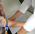
Tourniquet binding

Before injection

Inserted catheter ready for use
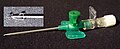
Catheter needle with new safety features.


Notes on cannulation and difficult cannulation[edit | edit source]
- Before using the cannula, it is a good idea to drive the mandrel into the lumen of the cannula several times so that the mandrel can slide freely out of the cannula during the actual cannulation.
- If, for any reason, the movement is stopped in the middle of the way (in the classic way), it is better to put the cannula back just under the skin and start the injection again. We always "back up" in the same direction as the puncture to prevent possible damage to the vein on the way back.
- If blood appears in the capillary cone only when "reversing", it is a sign that we have "passed" the vein, either by injecting too fast or too slowly, under stretching or overstretching the subcutaneous tissue, or when the needle is too inclined. We can still try to insert the cannula at the moment of the greatest return of blood, but the probability of success is already quite small.
- If the blood does not appear in the capillary cone at all and blood starts to flow out of the injection point after pulling the needle out, is again a sign that we have "driven through" the vein. Most often with excessive skin tension or cold acres, but the cause can also be a rapid puncture or hitting the vein at a completely wrong angle than its direction.
- In infants, toddlers or dehydrated children, it may happen that after cannulation blood will not flow out of the cannula after a drop of blood appears in our cone and we pull out the mandrel. If we are convinced that the injection was performed correctly, we do not pull out the cannula, but connect the connector and insert the cannula normally with the current injection. However, if we feel that the injection was performed incorrectly, it's a lottery whether to try to insert the cannula with a spray as in the previous case or to try to pull the cannula back slightly and insert it when blood begins to flow out of the cannula.
- If our skin "piles up" at the beginning of the injection, we will interrupt the injection and start again after the skin has been perfectly stretched !!!
- If we repeatedly and unsuccessfully move only in the subcutaneous tissue in an attempt to find the lumen of the vein, it is necessary to take into account the possibility of clogging the needle. Then it is better to either replace the cannula with a new one or rinse and then blow the needle with a good stream of air from the syringe. Rarely, a clogging of the needle can occur even after one attempt of "dry puncture".
- If we are still moving under the skin during the puncture in order to find the lumen of the vein, it is necessary not to deviate too much from the original axis of the puncture. Otherwise, there is a great risk that we will stab the vein at the "wrong" angle and despite the presence of blood in the signal chamber, we won't be able to insert the cannula.
- When cannulating in the dorsal area of the hands or feet, especially in the younger children, start the injection proximally enough (if the course of the vein allows) so that fixation of the signal chamber with the index finger of the right hand is possible just against the dorsum of the hand or foot. If we prick a lot distally, the signal chamber is "in the interspace" and the tip of the cannula can easily slip out of the lumen of the needle.
- If we choose to cannulate the cubital fossa and clearly do not see any suitable vein in this area, it is worth palpating the cubital area, as it is often possible to feel a strong venous trunk that may not be visible at all. If a "blue spot" shines in the cubital fossa, which indicates the presence of a vein, but its course is not visible, it is again appropriate to try to clarify the course of the vein by palpation. From a general point of view, the cubital area is a great place for cannulation, except in patients with markedly motor restlessness, because the fixation of the veins in it in a struggling child is far more complicated than in the dorsa area of the hand or foot. In these cases, it is advantageous if the nurses fix the forearms even distally and the cannulated patient has both hands at his disposal (it is possible to better stretch the skin and at the same time it is better to manipulate the cannula itself after injecting a vein). The mediolateral traction of the underside of the forearm also allows better vein fixation but the traction must be moderate so it doesn't close the lumen of the vein,
- It is also necessary to respect the laws of gravity, i.e. blood does not have to appear in the cone of the capillary if we prick "downhill" - this applies especially to the veins in the head area, when pricking the v. jugularis externa and children in the crib with a barricade.
- If we cannulate a child whose acres are very cold, we must first warm the periphery thoroughly. When cannulating the veins on cold limbs, blood usually does not appear at all in the cone of the capillary, the place of the puncture often does not even bleed = the image of a dry puncture, but after the limb is heated, the puncture can start to bleed. Either way, the vein is canceled and even after warming up we have to look for another place to cannulate.
- Cave! - when cannulating v. jugularis externa, do not rely on blood to appear in the cone (although this is often the case) → it is necessary to cannulate with an open sight. If I have the impression that I am in a vein, the needle should be pulled out.
- After removing the metal needle, we prevent blood spillage by compressing the vein in front of the cannula tip with the index finger of the free hand.
- If we inject a vein whose lumen is optimally visible only with minimal skin and subcutaneous tension, then it is necessary to take into account a small intraluminal pressure and therefore cannot rely on a drop of blood to appear in the capillary cone as a sign of proper penetration → better to puncture with an open sight and if I feel like I'm in a lumen of a vein, try to pull out the mandrel - if we're right, blood will start to flow out of the cannula and we can slide the cannula with the current flush into the final position.
- In children with thick skin, it is better to cannulate with a larger "start", i.e. about 0.5-1 cm (but too long a start and there is a risk of losing the ideal direction and angle of the cannula to the vein). We also prefer to cannulate "non-cooperating patients" with start-up.
- Analgesic sedation should be considered very carefully in non-cooperating patients. Motor restlessness, of course, reduces the success of cannulation, rapidly, especially in thinner veins and veins in the cubital fossa. In addition, the need to firmly fix the patient's limbs leads to excessive tension of the skin above the cannulation site, to a narrowing of the lumen of the veins and thus a greater probability of "passing" the vein. If the patient responds with a strong defensive reaction (usually flexion) during the initial injection → the cannulation is in the vast majority of cases unsuccessful (the skin wrinkles and twitches, a sharp movement changes the injection speed and the position of the vein). An alternative for these children are areas such as the head, the v. jugularis externa, but it is best not to regret the time and sedate the child adequately!
- If it is inevitable to cannulate a vein where "someone has already been", then we always prick proximal to the original puncture and only when the lumen is sufficiently filled. If the lumen is not clearly filled, it may happen that the blood does not appear in the capillary cone at all, or rather appears only when reversing. In this case, you can try the method with an open sight and a "leap forward".
- When cannulating the veins on the volar side of the forearm, we usually cannulate in the classic way, but we must choose a significantly tangential inclination of the needle, i.e. 10-15 ° and proceed at a slower speed within the "allowed" speed. When cannulating on the volar side of the wrist, we also choose a larger tangential slope, but we usually follow the "open visor" method.
- When cannulating thin veins (newborns, volar side of the wrist, etc.) using the "open visor" method, common cause of failure is a too deep of an initial injection. It is best to guide the initial cannula for a short distance and immediately check for blood in the signal chamber.
- When cannulating veins in the cubital fossa in newborns, we achieve the best subcutaneous tension using the mediolateral pull or a combination of mediolateral and proximodistal pull. The proximodistal pull alone is usually not able to ensure sufficient skin tension and cannulation is unsuccessful.
- If we have to cannulate thin veins, it is generally true that: we prick with an open visor, in very thin veins the presence of blood in the capillary cone is often not expected. We can choose a purple 26 G cannula and in the extreme case we can insert only the tip of the needle with the hole into the vein and try to insert the cannula afterwards - the probability of success is significantly lower. It should be noted that the thinnest veins simply cannot be technically cannulated, so it is important to differentiate them and not get into them at all! Often, under thorough subcutaneous tension, a "non-cannulable" thin vein may falsely appear strong enough (it looks like a green-blue stripe without a general lumen), but an attempt to cannulate is always unsuccessful. The thickness of the vein must therefore be carefully assessed, both before and after the subcutaneous tension. If the vein appears to be too weak, it must be ignored. When assessing the thickness of a vein the lumen is more important then it's width.
It should be noted that the thinnest veins simply cannot be technically cannulated, so it is important to differentiate them and not get into them at all!
- If no vein can be found in common injection sites, such as dorsal parts and cubital fossas, or it is difficult to estimate their course, it is advisable to look at these areas from the opposite side, ie from the position of a nurse. In the classic case, the doctor monitors the course of suitable veins in the direction "from fingers to arm", while the nurse "from arm to fingers". The nurse therefore takes the position of a doctor and strangles the relevant limbs, the doctor takes the position of a nurse. Unexpectedly often, can we find veins that completely escape our attention from the position of a doctor, or we get a better opportunity to estimate their direction.
- If we have 3-4 unsuccessful attempts, it is first necessary to calm down, find the best available vein again, reconsider the current procedure, especially the speed, and then choose between the classic technique or the technique with a "raised visor". As a last resort, before considering CVK or intraosseous entry, is cannulation of the v. jugularis or v.axillaris.
For children where the veins are not clear, it is better to have the nurses cannulate initially as to not unnecessarily lose self-confidence in a series of failed injections, when it is still not possible to figure out the right procedure (at least for now…)
- In order to better assess whether we are not making mistakes at the injection speed, it is necessary to repeatedly train the optimal injection speed (5-6 / 37 mm)! It happens that in time our ideal speed "falls out of hand" This is probably especially true in a situation where we have a series of failed attempts in a row, even for several patients. The logic of our mistake seems to be missing, but the common denominator is intrusion into a vein with the appearance of blood in the capillary cone, but with the impossibility of further inserting the cannula ("not possible" or "bulge"). The probability of bad speed is then very likely!
We always cannulate a vein whose lumen is sufficiently filled and at the same time the subcutaneous tissue is sufficiently taut. This note applies to all types of veins, but the most important thing is to keep this in mind when cannulating the v. saphena, vein in the head region and newborns.
In order to better assess whether we are not making mistakes at the injection speed, it is necessary to repeatedly train the optimal injection speed (5-6/37 mm)!
In chronic patients during cannulation of cubital fossa blindly it is necessary not to initially monitor the entry of the cannula under the skin, but to watch the cone of the capillary, and it seems that a larger injection angle is possible.
It also worked for me to "bend" the cannula when inserting into thin and superficial veins - before insertion (only if I have an antibiotic screen, or strictly sterilely), or when piercing the skin, gently push, bend the mandrel more vertically about 40 ° from the original horizontal direction, I won't pierce the vein through…
"Open visor" also known as "leap forward" procedure[edit | edit source]
We choose this technique where we cannulate thin veins, placed just below the surface or above level, in newborns, dystrophics or chronic patients. Simply everywhere where using the classical method, there is a risk that the blood will appear in the cone of the capillary only after passing through a vein.
The "open visor" procedure means that we follow the tip of the cannula during the injection and choose the depth of the injection according to our estimate of the vein placement. The movement must not be too slow. The Braun brand cannula needle is only 1-2 mm ahead of the plastic cannula, so we inject as if we only wanted to get a needle into the vein → a frequent failure is initially too deep of an injection or other too deep "leaps".
When trying with an open visor, we inject as if we only wanted to get a needle into the vein → a frequent failure is initially too deep of an injection or too deep other "jumps"
In a situation where a drop of blood does not appear in the cone of the capillary after the initial injection, we proceed with the "leap forward" method. If the vein is clearly visible, then follow the needle tip again and advance 2-3 mm forward, then stop and check for blood in the capillary cone. If blood appears, remove the needle and insert the cannula in a standard way. If blood does not appear, follow the same procedure. If we did not meet a vein along the way = no blood appeared in the cone of the capillary, we will slowly pull the cannula under the surface of the skin and try the same procedure in a slightly different direction. In the thinnest veins, blood may not appear in the capillary cone. If we think this could be the case, it's a good idea to try to pull out the mandrel - if blood rushes into the cannula, we'll try to insert the cannula with a spray and if the blood doesn't show up in the cannula then we reinsert the mandrel and continue on.
We can use the "leap forward" method in a situation where we do not indicate cannulation in the classic way, but do not see the lumen of the vein. Then we always move forward by 2-3 mm, but we only watch the end of the capillary
Common mistakes in cannulation[edit | edit source]
- The most common mistake is when I lead the puncture and the skin is not well stretched or the lumen of the vein is not clearly visible !!!
- Probably the second most common mistake is to start cannulating a vein that I am not convinced it is ideal enough (thin veins or veins where the direction of their course is not apparent) without devoting enough time to inspecting all the patient's available veins and selecting the best. (A typical situation is when I stab the child unbelievable amount of times, so that the final "success" comes from cannulating a vein with a good course and translucency, which I did not notice at all!)
- Too deep of an initial injection or jumps too long in the open visor method.
- Cannulation of veins that are so thin throughout the lumen that they cannot be technically cannulated at all (do not be fooled by the seemingly good thickness of the vein under tension).
A situation where I lead the puncture and the skin is not well stretched or the lumen of the vein is not clearly visible is the most common cause of failure.
Complications[edit | edit source]
- Cellulitis,
- Phlebitis,
- artifical artery cannulation,
- paravenous collection → risk of possible tissue damage,
- hematoma,
- thrombosis, eventually embolization of a part of the cannula.
All of these complications are an indication to remove the cannula.
If there are no signs of complications, we can leave the cannula in situ for longer period than the 72 hours that are commonly reported in literature.
References[edit | edit source]
Related Articles[edit | edit source]
External links[edit | edit source]
Source[edit | edit source]
- HAVRÁNEK, Jiří: Kanylace periferní žíly.