Parotid gland
The parotid gland , also called the parotid gland, is the largest salivary gland.
Anatomy and Histology[edit | edit source]
A purely serous tuboalveolar branched gland, which is located on the outside of the masseter muscle , extends cranially to the arcus zygomaticus , backwards to the auricle and passes to the proc. styloid . Caudally, it goes to the neck in front of the sternocleidomastoid muscle and the posterior belly of the digastricus muscle . The facial nerve (VII cranial nerve) comes to it , which branches in it and forms the parotideus plexus .
The outer surface of the gland together with the masseter muscle is covered by the surface sheet of fascia parotideomasseterica . The deep sheet of this fascia is involved in separating the gland from the pterygoideus medialis muscle and from all the surrounding muscles. The tractus angularis separates the gland from the trigonum submandibulare .
The duct of the parotid gland (ductus parotideus, Stensen's duct), 5-6 cm long, passes over the front edge of the masseter muscle and opens into the oral cavity at the height of the second upper molar.
The gland produces about one third of the total amount of saliva, the secretion is relatively rich in amylase.
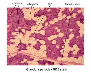
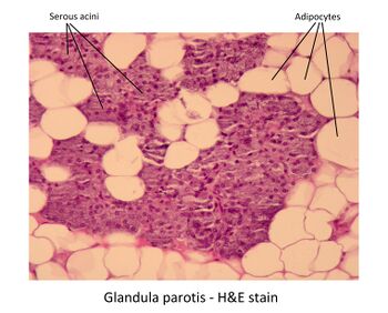
It contains serous alveoli, plasma cells, myoepithelial cells. We find inserted, annealed and interlobular ducts. It is typical for the parotid gland to contain many fat cells.
Vascular and nerve supply[edit | edit source]
The arteries to the gland area. temporalis superficialis, a. maxillaris, a. auricularis posterior and a. transversa faciei.
The veins come from the vv. parotideae.
The Nodi parotidei conduct lymph to the superficial and deep neck nodes.
Sympathetic fibers come along the arteries, parasympathetic fibers follow the path of the so-called Jacobson's anastomoses.
Histological images[edit | edit source]
Links[edit | edit source]
Related Articles[edit | edit source]
References[edit | edit source]
- MARTÍNEK, J. and Z. VACEK. Histological atlas. 1st edition. Grada, 2008. ISBN 978-80-247-2393-8 .
- KONRÁDOVÁ, V. , J. UHLÍK and L. VAJNER. Functional histology. 2nd edition. H&H, 2000. ISBN 80-86022-80-3 .


