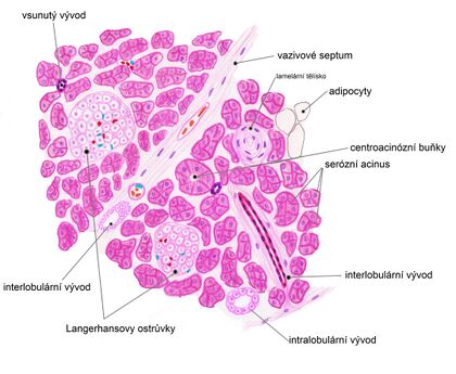
Pancreas (histological slide)
The pancreas is a retroperitoneal organ located in the epigastrium. Macroscopically, we divide the pancreas into three parts - caput, corpus, cauda.
We consider the pancreas to be a mixed gland and microscopically we can distinguish two parts - exocrine and endocrine.
Exocrine part[edit | edit source]
Secretory part[edit | edit source]
The pancreas is a purely serous gland in its structure (therefore it resembles the parotid gland and is often confused with it!). There are serous acini, which are formed by serous cells and represent the secretory component. The center of the acinus is formed by centroacinous cells, which are partly penetrating inserted ducts, see below. On the surface there is a basal lamina and the darker acinous cells are pyramidal in shape.
Acini cells synthesize digestive enzymes of pancreatic juice in the form of their precursors. It creates and stores enzymes such as trypsinogen, chymotrypsinogen, carboxypeptidase, ribonuclease.
Root part[edit | edit source]
The outlet component consists of inserted ducts, intralobular ducts, interlobular ducts and subsequently pancreatic ducts (ductus pancreaticus, ductus pancreaticus accessorius).
The inserted ducts branch and begin in the pancreatic acini, where they penetrate to the acini lumina . The lining of the intra-acinous part of the inserted duct is made up of centro-acinous cells. The cells do not contain zymogen granules and this causes them to stain lighter than normal acinar cells. The inserted ducts connect to the intralobular ducts (single-layered cubic epithelium), and then to the interlobular ducts (single-layered cylindrical epithelium) running in the fibrous septa, from which the pancreatic ducts arise. Pancreatic juice continues through the ductus pancreaticus to the duodenum.
![]() We do not find any annealed ducts in the pancreas.
We do not find any annealed ducts in the pancreas.
Endocrine part[edit | edit source]
In the pancreas we find the so-called islets of Langerhans , of which there are over a million (most in the tail). On the histological slide, the islets of Langerhans appear as light circular areas (resembling balls) that contrast with the surrounding darker colored exocrine tissue.
The islet itself consists of a beam-like assembly of several thousand epithelial cells, which are polygonal to round in shape. They are penetrated by a dense network of fenestrated capillaries, and the blood from these capillaries drains into the surrounding exocrine tissue, which is thus exposed to hormones from the endocrine part. The islets of Langerhans synthesize pancreatic hormones such as glucagon, insulin, somatostatin or pancreatic polypeptide.
See the Pancreatic Hormones page for more detailed information
Links[edit | edit source]
Related Articles[edit | edit source]
References[edit | edit source]
- L. CARLOS, Junquiera – JOSÉ, Carneiro – ROBERT O., Kelley. Základy histologie. 1. edition. Jinočany : H & H 1997, 1997. 502 pp. ISBN 80-85787-37-7.
- RENATE, Lüllmann-Rauch. Histologie. - edition. Grada Publishing a.s., 2012. 556 pp. ISBN 9788024737294.
- JAN, Balko – ZBYNĚK, Tonar – IVAN, Varga. Memorix histologie. 1. edition. Triton, 2016. ISBN 9788075530097.

