POSITRON EMISSION TOMOGRAPHY (PET)
- Previous chapter: 6.5 SINGLE-PHOTON EMISSION COMPUTED TOMOGRAPHY (SPECT)
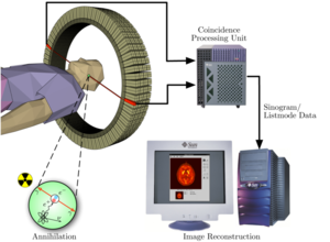
Positron emission tomography, also called PET imaging or a PET scan, is a type of nuclear medicine imaging that produces a three-dimensional image or picture of functional processes in the body. This system detects pairs of gamma rays emitted indirectly by a positron-emitting radionuclide, which is introduced into the body on a biologically active molecule.
PET technology can be used to trace the biologic pathway of any compound in living humans (and many other species as well), provided it can be radiolabeled with a PET isotope. PET is both a medical and research tool. It is used heavily / frequently in clinical oncology (medical imaging of tumors and the search for metastases), and for clinical diagnosis of certain diffuse brain diseases such as those causing various types of dementias. PET is also an important research tool to map normal human brain and heart function.
To conduct the scan, a short-lived radioactive tracer isotope is injected into the living subject (usually into blood circulation). Usually short half-life isotopes are used like carbon-11 (~20 min), nitrogen-13 (~10 min), oxygen-15 (~2 min), fluorine-18 (~110 min)., or rubidum-82(~1.27 min). These radionuclides are incorporated either into compounds normally used by the body such as glucose (or glucose analogues), water, or ammonia, or into molecules that bind to receptors or other sites of drug action.
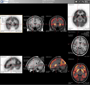
The tracer is chemically incorporated into a biologically active molecule. As the radioisotope undergoes positron emission decay (also known as positive beta decay), it emits a positron, an antiparticle of the electron with opposite charge. The most significant fraction of electron-positron decays result in two 511 keV gamma photons being emitted at almost 180 degrees to each other; hence, it is possible to localize their source along a straight line of coincidence. Nearly same like in SPECT imaging also a suitable detector has to be used (PMT or avalanche diode) Similarly to SPECT imaging we also have to use a suitable detector. Then the acquired data is processed and a functional image is reconstructed. Because of lower resolution than in other techniques it is often combined with other imaging methods like CTs or MRIs. The CT or MRI produce the anatomical image and the PET gives the functional image. Usually for that are modern CTs or MRIs combined with PET detectors. Modern CTs or MRIs are usually combined with PET detectors.
Links[edit | edit source]
Next chapter: 6.7 MAGNETIC RESONANCE IMAGING (MRI)
Back to Contents


