Of cerebrospinal fluid
Cerebrospinal fluid (also liquor , liquor cerebrospinalis , cerebrospinal fluid - CSF) is a clear, colorless liquid. It has an identical qualitative but different quantitative composition compared to plasma.
Formation, circulation and resorption of cerebrospinal fluid
Cerebrospinal fluid is produced by active secretion by cells of the choroid plexus and ependyma of individual cerebral ventricles (50–70%). Another portion is created by ultrafiltration of blood plasma by choroidal capillaries. The volume of cerebrospinal fluid in an adult is about 120-180 ml. It is produced at a rate of about 500–600 ml per 24 hours . CSF secretion persists even when outflow is prevented by obstruction, then it accumulates and increases intracranial pressure.
The cerebrospinal fluid is located ``intracerebrally (20%) in the region of the two lateral ventricles, the third and fourth ventricles and connections between the ventricles, and extracerebrally (subarachnoidally) (80 %) in the space between the pia mater and the arachnoid on the surface of the brain and spinal cord.
The Circulation of CSF begins in the lateral ventricles, continues through the third and fourth ventricles, and continues to flow into the subarachnoid space. Part of the cerebrospinal fluid surrounds the brain stem and spinal cord.
The ``resorption of the cerebrospinal fluid takes place through the arachnoid villi of the ``Pacchionian granulations (granulationes arachnoideae), through which it passes into the large intracranial venous sinuses. In this way, the direct transfer of cerebrospinal fluid into the venous circulation is ensured.
Function of cerebrospinal fluid
Cerebrospinal fluid performs several functions:
- Mechanical - surrounds the brain and spinal cord from all sides and thus protects them from shocks, changes in pressure and temperature.
- Homeostatic - ensures optimal environment for CNS cells (constant ion composition, pH and osmolarity).
- Metabolic - ensures removal of catabolism products (eg lactate, CO2) and supplies various bioactive substances to brain cells.
- Protective - participates in protection against pathogenic microorganisms.
Barriers
The composition of the cerebrospinal fluid affects the barrier system. The concept of the blood-brain barrier includes the interface between the blood, the brain and the cerebrospinal fluid, which allows some substances to pass in both directions or only in one direction and may restrict the passage of other substances. The blood-encephalic barrier provides an optimal environment for brain function, protects the brain from harmful substances and enables the brain to be supplied with substances needed for its metabolism. We distinguish between the "blood-encephalic", "encephalo-CSF" and "blood-CSF barrier".
Blood-brain barrier
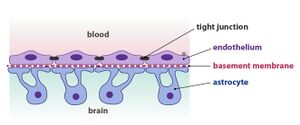
The blood-brain barrier (in the narrower sense) creates a transition between brain capillaries and brain tissue . The morphological basis of the blood-brain barrier is a continuous layer of cerebral capillary endothelium on the blood side, a basement membrane and a layer of astrocytes on the brain side . Brain capillary endothelium differs from endothelium in other locations in that it is free of fenestrations and endothelial cells are connected by tight junction . Astrocyte protrusions are attached to the basement membrane together with pericytes ( microglial cells ) (Fig. 1).
Blood-liquid barrier
The blood and cerebrospinal fluid barrier separates blood and cerebrospinal fluid.
It is formed by the epithelium of the choroid plexus, which secretes cerebrospinal fluid. Epithelial cells are connected by tight junctions that are more permeable than the tight junctions in brain capillaries. On the side facing the cerebrospinal fluid, they create microvilli that significantly increase the epithelial surface. diffusion, facilitated diffusion and active transport into the CSF, but also transport from the CSF into the circulation take place in the choroid plexus.
Another part of the blood-liquid barrier are the capillaries of the pia mater, which are fenestrated and resemble capillaries in other locations (Fig. 2).
The blood-liquid barrier is more permeable and enables the transfer of proteins from the plasma to the liquid via pinocytosis or specific carriers. A disorder of the blood-cerebrospinal fluid barrier is manifested by increased concentrations of proteins in the cerebrospinal fluid (further see albumin and immunoglobulins in the cerebrospinal fluid).
Cerebrospinal fluid barrier
The essence of the CSF barrier is a layer of glial fibers on the surface of the brain and the ependyma of the ventricles. This barrier is more permeable than the blood-liquid barrier. Penetration of substances takes place through the intercellular gaps in the glia layer and the gaps between the ependyma of the ventricles. Protein-sized substances can diffuse in both directions.
Cerebrospinal fluid examination
Cerebrospinal fluid testing is one of the basic methods that contribute to the diagnosis of neurological diseases. The cerebrospinal fluid is most often collected by lumbar puncture (3×5 ml, between L4–L5 or S1), the suboccipital approach is less common. The cerebrospinal fluid needs to be transported to the laboratory as quickly as possible, as the cells gradually break down, the glucose concentration decreases and the lactate increases.
- The basic examination of cerebrospinal fluid includes the performance of these analyzes
- assessment of the appearance of cerebrospinal fluid,
- quantitative determination of total protein,
- quantitative determination of lactate,
- qualitative and quantitative cytological examination,
- cerebrospinal fluid spectrophotometry.
- Further examination of cerebrospinal fluid includes these determinations
- determination of IgG, IgA, IgM, albumin in plasma and cerebrospinal fluid with estimation of intrathecal immunoglobulin synthesis and determination of blood-brain barrier disorder,
- isoelectric focusing for the detection of IgG oligoclonal bands,
- some other tests (e.g. determination of other proteins in cerebrospinal fluid and serum, specially stained cytological preparations).
- Cerebrospinal fluid is mainly examined in these diseases
- suspected acute neuroinfection,
- suspected subarachnoid hemorrhage,
- demyelinating disease,
- malignant infiltration of meninges.
- Contraindications to consumption
- finding of intracranial expansion process,
- Intracranial hypertension,
- local infection at the puncture site,
- some coagulopathies,
- we take 1–2 ml separately due to artificial blood contamination.
Links
related articles
- Liquor examination
- Biochemické vyšetření mozkomíšního moku
- Bílkoviny v mozkomíšním moku
- Spektrofotometrie mozkomíšního moku
- Cytologické vyšetření mozkomíšního moku
- Liquor syndromes
References
- ADAM, P, et al. Cerebrospinal fluid cytology (CD-ROM). 1. edition. Prague : SEKK, 2000.
- AMBLER, Z – BEDNAŘÍK, J – RŮŽIČKA, E. Clinical neurology – general part. 1. edition. Prague : Triton, 2004. ISBN 80-7254-556-6.
- GLOSOVÁ, L. Cytological atlas of cerebrospinal fluid. 1. edition. Prague : Galén, 1998. ISBN 80- 85824-70-1.
- KALA, M. – MAREŠ, J. Lumbar puncture and cerebrospinal fluid. 1. edition. Prague : Galén, 2008. ISBN 978-80-7262-568-0.
- MASOPUST, J. Clinical Biochemistry. Requesting and evaluation of biochemical examinations I. and II. part. 1. edition. Prague : Karolinum, 1998. ISBN 80-7184-650-3.
- NEVŠÍMALOVÁ, S – RŮŽIČKA, E – TICHÝ, J, et al. Neurology. 1. edition. Galen, 2005. ISBN 80-7262-160-2.
- SCHNEIDERKA, Peter. Chapters in Clinical Biochemistry. 2. edition. Karolinum, 2004. ISBN 80-246-0678-X.
- SEAGULL, J, et al. Clinical Biochemistry. 1. edition. Galen – Karolinum, 1999. ISBN 80-7262-023-1.
- ŠTERN, P, et al. General and clinical biochemistry for bachelor's fields of study. 1. edition. Prague : Karolinum, 2005. ISBN 978-80-246-1025-2.
- WINTER, T, et al. Laboratory diagnostics. 1. edition. Prague : Galen – Karolinum, 2002. ISBN 80-7262-201-3.


