Methemoglobin
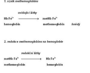
Methemoglobin (metHb; also hemiglobin or ferihemoglobin[1]) is characterized by the presence of trivalent iron, which is formed by the oxidation of divalent iron in hemoglobin[2]. Methemoglobin loses its ability to reversibly bind oxygen. In its place, FeIII+ binds a water molecule with a sixth coordination bond. The color of methemoglobin is chocolate brown.
Methemoglobin is also present physiologically in small amounts in erythrocytes (about 1–3% of the total concentration of hemoglobin[3]). This is mainly due to the effect of nitrites, which arise from nitrates contained in food. The reverse reduction of methemoglobin to hemoglobin is primarily ensured by the NADH-dependent cytochrome-b5 reductase (also methemoglobin reductase). A minor role is played by NADPH-dependent methemoglobin reductase, which is dependent on the supply of NADPH from the pentose cycle and on the presence of another electron carrier (e.g. flavin).[4] Non-enzymatic mechanisms include the action of glutathione and ascorbic acids.
 An increased concentration of methemoglobin in the blood is referred to as 'methemoglobinemia. The causes are various:
- Hereditary methemoglobinemia is usually caused by a congenital defect in NADH-dependent methemoglobin reductase or the presence of abnormal hemoglobin M.
- Acquired methemoglobinemia is the most common form of methemoglobinemia. It can be caused by the action of oxidizing substances[5]:
- poisoning by certain substances (nitrobenzene, aniline and its derivatives - e.g. some dyes),
- under the influence of certain drugs (local anesthetics – benzocaine, also phenacetin, sulfonamides),
- increased content of nitrates and nitrites in water and food.
Newborns are especially sensitive to the increased content of these substances due to the immaturity of the reduction systems and the increased proportion of fetal hemoglobin, which is more easily oxidized. Methemoglobinemia manifests as cyanosis with a characteristic gray-brown hue and hypoxia.
| Methemoglobin values | Symptoms |
|---|---|
| 0-2% | normal value |
| < 10% | cyanosis |
| < 35% | cyanosis and other symptoms (headache, shortness of breath) |
| 70% | lethal concentration |
Part of the therapy of acquired methemoglobinemia is the administration of some reducing agents - methylene blue or ascorbic acid.
Links[edit | edit source]
External links[edit | edit source]
Source[edit | edit source]
- ↑ ŠVÍGLEROVA, Jitka. Hemoglobin [online]. Last revision 2009-02-18, [cit. 2010-11-11]. <https://web.archive.org/web/20160416205421/http://wiki.lfp-studium.cz/index.php/Hemoglobin>.
- ↑ ŠVECOVÁ, D and D BÖHMER. Congenital and acquired methemoglobinemia and their treatment. Journal of Czech doctors. 1998, vol. 137, pp. 168-170, ISSN 1803-6597.
- ↑ RICHARD, Alyce M, James H DIAZ, and Alan David KAYE. Reexamining the risks of drinking-water nitrates on public health. Ochsner J [online]. 2014, vol. 14, no. 3, pp. 392-8, also available from <https://www.ncbi.nlm.nih.gov/pmc/articles/PMC4171798/?tool=pubmed>. ISSN 1524-5012.
- ↑ XU, F, K S QUANDT and D E HULTQUIST. Characterization of NADPH-dependent methemoglobin reductase as a heme-binding protein present in erythrocytes and liver. Proc Natl Acad Sci U S A [online]. 1992, vol. 89, no. 6, pp. 2130-4, also available from <https://www.ncbi.nlm.nih.gov/pmc/articles/PMC48610/?tool=pubmed>. ISSN 0027-8424.
- ↑ CORTAZZO, Jessica A, and Adam D LICHTMAN. Methemoglobinemia: a review and recommendations for management. J Cardiothorac Vasc Anesth [online]. 2014, vol. 28, no. 4, pp. 1043-7, also available from <https://www.ncbi.nlm.nih.gov/pubmed/23953868>. ISSN 1053-0770 (print), 1532-8422.
