Meninges


Meninges (sg. meninx) are membranous layers covering and protecting central nervous system (brain and spinal cord).
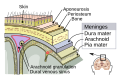
The meninges consist of 3 fibrous layers (from superficial → deep):
- Dura Mater
- Arachnoid
- Pia Mater
Dura Mater[edit | edit source]
It is derived from ectomeninx.
- Encephalic dura mater: it is closely attached to the periosteum. Coalescence takes place and veins lose their external layer to form sinuses. It has the following infoldings:
- Falx Cerebri – located sagitally in longitudinal fissure of cerebrum, starting from Crista galli and frontalis and ending at the internal occipital protruberance. Beneath it is corpus callosum.
- Tentorium Cerebelli – overlies the cerebellum and thus separating it from the occipital lobes. Attached on the internal occipital protuberance and continues along the sulcus of transverse sinus as far as the upper edge of petrous bone. Anterior margin is free, forming an aperture the tentorial incisure under which the brainstem passes from the posterior cranial fossa to the central fossa.
- Falx Cerebelli – extension of falx cerebri.
- Diaphragm sellae – forms the roof of the hypophysial fossa.
- Spinal dura mater: No coalescence takes place, so veins are left intact, forming the internal venous plexuses, in the epidural space. It forms the sac of spinal dura mater. External to it, there are the internal venous plexuses and external to them, the endorachis. It reaches as far S2 vertebra – it then continues as thread-like extensions, terminal filum of the spinal dura mater, up to the coccygeal bone. It is fixed on the spinal cord by the roots of spinal nerves on which it extends along them, up to the intervertebral foramina.
Arachnoid[edit | edit source]
It is a thin avascular membrane, derived from the endomeninx.
- Encephalic arachnoid: in the superior sagittal sinus, the arachnoid gives off extensions into the superior sagittal sinus and into the diploic vessels in the periosteum. These extensions are called arachnoidal granulations. They are involved in draining the CSF from the subarachnoid cavities into the venous system. The encephalic arachnoid spreads over the whole brain, but not reaching the deeper fissures. Pia mater follows them closely, thus leading to a detachment of the 2 membranes, forming enlarged subarachnoid cavities/cisternae filled with CSF:
- Cerebellomedullar cistern (below cerebellum),
- Quadrigeminal cistern (above cerebellum),
- Cisterna of lateral fossa (above insula),
- Chiasmatic cisterna (around optic chiasm),
- Interpenducular cisterna (over the synonymous fossa).
- Spinal arachnoid: the trabeculae are organized in a more robust form, located alongside of the spinal cord, forming a system of flat denticulate extensions, called denticulate ligament. It follows the spinal roots and spinal nerves, blending as a thin layer of flattened cells with perineurium and follows the peripheral nerves until they reach their destination.
Pia Mater[edit | edit source]
It is derived from endomeninx. It adheres closely on the surface of brain and spinal cord. On its surface run larger vessels among the trabeculae (in the subarachnoid space), which penetrate the cerebral tissue.
Innervation & Vasculature[edit | edit source]
The cranial meninges are innervated by the middle meningeal nerve and the nervus spinosus. They are supplied by the:
- Middle meningeal artery.
- Meningeal branches of the ascending pharyngeal artery.
- Accessory meningeal artery.
- Branch of anterior ethmoidal artery.
- Meningeal branches of vertebral artery.
The spinal meninges are supplied by:
- Anterior spinal artery (in subarachnoid space).
- Paired posterior spinal arteries with the corresponding spinal veins (in subarachnoid space).
The spinal arteries form anastomoses known as the vasocorona of the spinal cord.
- Internal vertebral venous plexuses (in epidural space).
Pathology[edit | edit source]
Hemorrhage:
- Subarachnoid hemorrhage is acute bleeding under the arachnoid. Most often it is from a ruptured aneurysm, they tend to be located in the circle of Willi. Arterialvenous malformation (congenital) is the second most common cause of subarachnoidal hemorrhage. The sudden change into bleeding from these cause can be due to trauma or spontaneously.
- Subdural hematoma is a hematoma located in a separation of the arachnoid from the dura mater. The small veins which connect the dura mater and the arachnoid are torn, usually during an accident, and blood can leak into this area. The collection of blood is slow (thus no mass-effects in early stages) due to the low-pressure system of the veins and thus can be easily missed in the patient's (probably asymptomatic) clinical picture.
- Epidural hematoma similarly may arise after an accident or spontaneously. In epidural hematoma most commonly the middle menginal artery is involve, however also other arteries and in rare cases even veins may be the origin of the hematoma.
Other medical conditions:
- Meningitis (fungal, bacterial, or viral infection).
- Meningiomas arising from the meninges or from tumors formed elsewhere in the body which metastasize to the meninges.
Summary of Cranial & Spinal Menignes & Layers[edit | edit source]
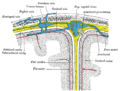
Diagrammatic representation of a section across the top of the skull
Meningeal Layers


| Cranium | Spinal cord |
|---|---|
| — | Endorachis |
| (Sinuses) | Epidural space |
| Dura mater | Dura mater |
| Subdural space | Subdural space |
| Arachnoid | Arachnoid |
| Subarachnoid space | Subarachnoid space |
| Pia mater | Pia mater |
Links[edit | edit source]
Sources[edit | edit source]
- Lecture Notes: Prof. MUDr. Jaroslav Pokorný DrSc.