Mechanics of breathing
Breathing (respiration) belongs to the basic processes during which gas exchange takes place in the organism. Oxygen is taken in during respiration, and carbon dioxide, which is produced as a product of oxidation processes, is on the contrary eliminated. We distinguish breathing into:
- internal (tissue respiration) – exchange O2 a CO2 between blood and tissues;
- external (pulmonary respiration) – difffusion of O2 and CO2 From the air into the blood
External Breathing[edit | edit source]
External respiration consists of 4 basic processes:
- Pulmonary ventilation
- Distribution
- Diffusion
- Perfusion
1. Pulmonary ventilation[edit | edit source]
Pulmonary ventilation refers to the exchange of air between the lungs and the outside environment. It is possible due to the pressure difference between the atmosphere and the alveoli. The pressures inside the chest change due to the movement of the respiratory muscles (so-called respiratory movements). The flow of air in the lungs is periodically repeated as inhalation (inspirium) and exhalation (expirium). Ventilation is subject to central control based on various parameters (e.g. blood pH, concentration CO2, concentration O2).
Inspirium[edit | edit source]
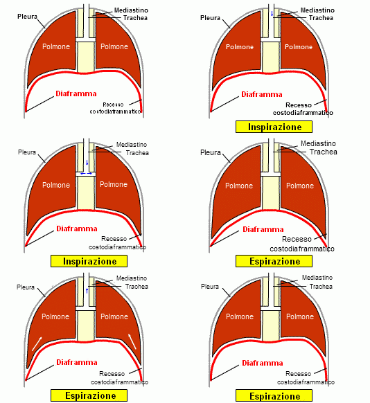
It is always an active plot. Air is drawn into the lungs thanks to the pressure drop in the lungs, created by the action of the inspiratory muscles Diaphragm descends about 1 cm. The ribs are raised using the external intercostal muscles.
Expirium[edit | edit source]
Under physiological conditions at rest, this is a passive event. The pressure drop is directed out of the lungs, air is expelled from the lungs. Contraction of the alveoli takes place due to the effect of elastic fibers and surface tension (the so-called retraction force of the lungs to the hilum).
These events occur due to negative pleural pressure (this pressure is lower than atmospheric pressure). Negative pleural pressure changes during breathing. It has the highest value during exhalation, and the lowest during inhalation. The highest value is around −2 cm H2O, the lowest −8 cm H2O[1].
Pneumothorax occurs when negative intrathoracic pressure is violated. In pneumothorax, the affected part of the lung is completely or partially excluded from respiratory activities.
Minute ventilation[edit | edit source]
Minute ventilation is a physiological parameter that varies depending on the load in the range 6–180 liters/min[1]. It is determined as the volume of inhaled or exhaled air (not their sum). Roughly speaking, it can be defined as the product of respiratory volume (VT) and respiratory frequency (f).
MV = VT × f
During exercise, the value of minute ventilation changes due to changes in respiratory volume and respiratory rate.
Values[1][edit | edit source]
| Parameter | Value |
|---|---|
| Minute ventilation (MV) | 5–8 liters |
| Maximum minute ventilation (MMV) | 200 liters |
| Tidal volume objem (VT) | 400–500 ml |
| Respiratory rate (f) | 12–16/min |
2. Distribution[edit | edit source]
During distribution, the inhaled air is mixed with the air that remained in the airways and lungs after the previous exhalation. There is no respiratory epithelium in the upper and lower respiratory tracts, including the bronchioles, so gas exchange cannot take place in them. This space is referred to as anatomical dead breathing space (VD=150 ml).
3. Diffusion[edit | edit source]
Gas exchange itself (O2, CO2) takes place in the pulmonary vaults (alveoli). The total surface of the alveoli is 100 m2[2]. Adjacent to the alveoli is a dense network of capillaries.
Diffusion is the passage of oxygen and carbon dioxide across the alveolocapillary membrane. Oxygen passes from the alveoli to the pulmonary capillaries, carbon dioxide passes from the capillaries to the the alveoli.
Rate of diffusion[edit | edit source]
The diffusion rate is based on Fick´s law. The volume of gas that passes through the membrane per unit time is directly proportional to the difference in partial pressures on both sides of the membrane (P1, P2), the membrane area (A) and the diffusion constant (K) and inversely proportional to membrane thickness (T).
The rate of diffusion of CO2 through the alveolocapillary membrane is 20,6 times greater than the rate of diffusion of O2.
Diffusion constant[edit | edit source]
The diffusion constant is dependent on the composition of the membrane and the type of gas passing through.
Partial pressures of breathing gases[edit | edit source]
The partial pressures of O2 and CO2 depend on how much of these gases are physically dissolved in the blood. The partial pressures of gases in arterial blood are different from those on venous blood. The partial pressure of O2 in arterial blood is 12,7 kPa, in venous blood it is 5,2 kPa. The partial pressure of CO2 in arterial blood is 5,2 kPa, in venous blood it is 6,13 kPa.
4. Perfusion[edit | edit source]
Perfusion is the flow of blood through the pulmonary capillaries, It is important for maintaining the pressure gradient for oxygen and carbon dioxide. These gases are carried in the blood in different forms.
Oxygen[edit | edit source]
Oxygen exists in the blood in two forms. Most of the volume of O2 In arterial blood is chemically bound to hemoglobin ( 197 ml O2 / 1 l of blood ), the rest is physically dissolved in the blood ( 3 ml O2 / 1 l of blood ). However, physically dissolved oxygen is very important in the blood, as it creates a partial pressure and thus allows diffusion.
Carbon monoxide[edit | edit source]
Carbon dioxide exists in the blood in three forms. Either it physically dissolved in it ( 30 ml / 1 l of blood ), or it is bound in the from of bicarbonates or carbamine compounds ( 520 ml / 1 l of blood ).
Respiratory resistances[edit | edit source]
Elastic lung resistance[edit | edit source]
It ensures the flexibility of the lungs. During breathing, this resistance is overcome thanks to the respiratory muscles. (Compliance), or the change in lung volume as a function of pressure change, is an indicator of the force that must be exerted on the lung tissue to cause the lungs to expand. The more flexible the lung, the greater the compliance.
Inelastic tissue resistance[edit | edit source]
Viscous resistance arising from the friction of lung tissue, chest, respiratory muscles and organs of the abdominal cavity.
Airway current resistance[edit | edit source]
It is determined by the pressure required to overcome airway resistance. We distinguish between laminar flow, turbulent flow and transitional floe. Flow resistance is characterized as the value of the alveolar pressure that ensures the flow of 1l of air per second.
The total work of breathing is determined by the mechanical effort expended to overcome mechanical resistances.
Links[edit | edit source]
Related articles[edit | edit source]
- Lung
- Diffusion
- Equation of state of gases
- Daltońs Law
- Henry´s Law
- Fick´s Laws: • 1. Fick´s Law
- Cheyneo-Stokes respiration
- Biot´s Breath
- Kussmaul respiration
- Nervous regulation of breathing
- Chemical regulation of respiration
References[edit | edit source]
Used literatuure[edit | edit source]
- HRAZDIRA, Ivo – MORNSTEIN, Vojtěch. Lékařská biofyzika a přístrojová technika. 1. edition. Brno : Neptun, 2001. ISBN 80-902896-1-4.
- NAVRÁTIL, Leoš – ROSINA, Jozef. Medicínská biofyzika. 1. edition. Praha : GRADA, 2005. ISBN 80-247-1152-4.


