Lichen ruber planus
From WikiLectures
Lichen ruber planus belongs between erythemato-papulo-squamous dermatosis, whose characteristic characters are :
- itching flat reddish brown papules around 1& thinsp;mm in diameter, waxy shiny,
- typical histopathological finding,
- disabilities mucous membranes, hair and nails.
Etiology[edit | edit source]
Etiology the disease is unclear. Participation is considered cytotoxic T-lymphocytes directed against antigens in the area basal membranes. Illness has connection with chronic hepatopathies (hepatitis C andhepatitis D) and administration drugs (beta - blockers).
Clinical image[edit | edit source]
typical for lichen planus symmetrical sowing itchy, flat,shiny,polygonal,reddish-brown papule. On the surface papule they can be sometimes visible Wickham's stria – whitish drawing, which is conditional hypergranulosis. After healing papule they persist hyperpigmentation.
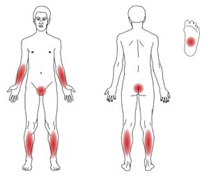
Predilection localization are :
- volar parties wrist,
- cross landscape,
- insteps, ankles.
Clinical forms[edit | edit source]
- Exanthematic form
- Acts massive acute sowing petty cash papule mainly on hull which can go to erythroderma. He can be present asymptomatic whitish reticulated venation on buccal mucosa (up to half sick). At 10 % sick people occur and changes on nails
- Lichen planus annularis
- Speeches sometimes they can be grouped into rings. This form often affects genitalia.
- Lichen planus mucosae
- Manifests as painful erosions and scarring, especially around cavities oral and anu.
Lichen planopilaris
- The emergence is characteristic follicularly bound and often confluent pointed hyperkeratotic red ones papule. He can lead to scarring alopecia.
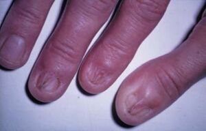
Lichen unguium
- Running out to thinning disc, deformation disk. Subungual hyperkeratosis they can lead up to total loss nails.
Lichen palmoplantaris
- It is diffuse reddish-brown hyperkeratosis face and palm, sometimes with ulcerations
- Lichen planus verrucosus
- They arise verrucous elevated reddish brown bearings, often on shins.
Histopathological finding[edit | edit source]
- Acanthosis, hypergranulosis, orthohyperkeratosis epidermis,
- mononuclear striped infiltrate in the upper corium penetrating into the lower ones parts epidermis, dermo- epidermoid the junction is not sharp, forming " teeth saws ",
- vacuolar degeneration of keratinocytes in the basal layer, clusters cytoid corpuscles – Civatte corpuscles,
- shedding of melanin to the corium – here the melanin pigment is absorbed by macrophages,
- direct immunofluorescence demonstrates immunopositivity IgM and IgG.
Differential diagnosis[edit | edit source]
Therapy[edit | edit source]
- Local – corticosteroids, application of immunomodulation (tacrolimus, pimecrolimus), disinfectant lavages in the disabled mucous membranes, local anesthetics.
- Total treatment in extensive forms – corticosteroids or retinoids.
Links[edit | edit source]
Related articles[edit | edit source]
References[edit | edit source]
- STORK, George, et al. Dermatovenereology. 1. edition. Prague : Galen, 2008. 502 pp. ISBN 978-80-7262-371-6.


