Larynx
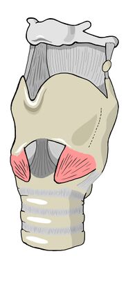
The larynx (Latin larynx ) is an unpaired organ, it belongs to the respiratory system. Air passes through it into the trachea and then into the lungs , thus serving for respiration and further for phonation .
It is stored in the anterior region of the neck. The wall of the larynx is strengthened by cartilage, small joints, ligaments and membranes. Cartilage is controlled by 7 pairs of muscles. The surface is covered by adventitia and the cavity is lined with mucous membrane, which is attached with the help of submucosal ligament ( histological preparation ).
The shape of the larynx[edit | edit source]
The larynx is hollow, flattened behind. Its length is 5 cm. The cavity of the larynx, cavitas laryngis , is narrowed in the middle by two pairs of cilia, so it takes the shape of an hourglass. Narrowings and folds divide the laryngeal cavity into three parts: vestibulum laryngis, cavitas laryngis intermedia and cavitas infraglottica.
Vestibulum laryngis – vestibule, starts from the pharyngeal cavity through the entrance, aditus laryngis . The entrance is bounded from the front by the unpaired laryngeal valve, epiglottis , and on the sides by paired eyelashes, plicae aryepiglotticae . The eyelash is thickened in two bumps – medially tuberculum corniculatum and laterally tuberculum cuneiforme , both bumps are conditioned by cartilages of the same name. At the back, the aditus laryngis is closed by a fold sunk between the vocal cartilages, the plica interarytenoidea . The cavity of the vestibule narrows caudally and at the end there is a fold, plica vestibularis , which with the bilateral fold closes the slit, rima vestibuli. Its edges are rounded and deep red.
Cavitas laryngis intermedia - the middle part of the larynx, it expands to the sides and forms the ventriculus laryngis , which can further expand both ventrally and laterally into the sacullus laryngis . The lower border of the cavity is formed by the vocal folds, plicae vocales , which squeeze a gap between them, rima glottidis . The mucosa of the plicae vocales is white and covered by a stratified squamous epithelium . The front section of the rima glottidis is supported by a ligament, the ligamenta vocalia , it is referred to as the pars intermembranacea . In the posterior compartment, the rima is bounded by the vocal cartilage, cartilago arytaenoidea, and is the pars intercartilaginea .
Cavitas infraglottica – the lower part of the larynx, which smoothly passes into the trachea .
Location and syntopy[edit | edit source]
The larynx is stored in the visceral neck space. It is palpable and visible due to the arching of the skin by the laryngeal cartilages - prominentia laryngis (more visible in men, like a "bite"). Ventrally, only the superficial and middle cervical fascia lie in the midline. If formed, the lobus pyramidalis of the thyroid gland may also lie ventrally . Laterally are the lobes of the thyroid gland. The neurovascular bundle of the neck lies dorsolaterally . Dorsally lies the pharynx . Cranially, there is the tongue and uvula , on which the larynx is suspended.
In men, the larynx projects onto the bodies of vertebrae C 5-6 , in women it is half a vertebra higher. In newborns, it is placed more cranially than in adults, in front of the C 3-4 body , this ensures the possibility of swallowing during sucking.
The larynx is movable, therefore its position changes during swallowing, phonation, coughing, etc.
Cartilages of the larynx[edit | edit source]
Forms the skeleton of the larynx. Cartilago thyroidea, cricoidea and epiglottis are unpaired , the others: cartilago arytenoidea, corniculata, cuneiformis, are paired . Only the base for the epiglottis is elastic, otherwise the others have a hyaline base.
- Cartilago thyroidea − thyroid cartilage, is the most voluminous. There is a distinct prominence of the laryngis on the front surface of the neck (it is more pronounced in men than in women after puberty). Cartilage consists of two plates: lamina dextra et lamina sinistra . They are attached to each other at an angle of 90° and at the point of connection there is a notch above and below: incisura thyroidea superior et inscisura thyroidea inferior . From the dorsal edges of the discs, projections extend up and down on each side: cornu superius et cornu inferius . The cornu inferius have an articular surface for connection with the cartilago cricoidea. There is a raised oblique edge, linea obliqua , on the outer surface of the discs, which is the site of attachment and origin of the infrahyoid muscles . Dorsally from the linea obliqua, muscle bundles of the inferior constrictor pharyngis muscle begin . Ventrally from the linea obliqua there is usually an opening, the foramen thyroideum , for the r. internus n. laryngei superioris and the connected vessels (if they go through the membrana thyroidea, then the foramen is absent).
- Cartilago cricoidea – annular cartilage. It has an arch in front, arcus cartilaginis cricoideae , and a plate behind, lamina cartilaginis cricoideae . On the upper surface of the disc are paired articular fossae, facies articulares arytaenoideae , for connection with the cartilago arytaenoidea. On the back surface of the disc there are paired pits for the beginnings of muscles. On the side, at the point of transition of the arch into the disc, there is a paired articular fossa, facies articularis thyroidea .
- Cartilago arytaenoidea – vocal cartilage, paired. It has the shape of a pyramid with the base, basis , turned caudally and the top, apex , directed cranially. On the base there is an articular surface, facies articularis , for connection with the cartilago cricoidea. The processus vocalis pro lig extends forward from the base . vocale and laterally the processus muscularis for the attachment of the muscles of the larynx.
- Epiglottis – laryngeal valve, unpaired cartilago epiglottica, as the only one of elastic cartilage. Cranially, the epiglottis is wide, blade-shaped, extending caudally into a petiole , which is connected by a syndesmosis to the posterior surface of the cartilago thyroidea.
- Cartilago corniculata – small paired cartilage connected by syndesmosis with the tip of cartilago arytaenoidea. In the aditus laryngis, it conditions the arching of the mucous membrane into a tubercle, tuberculum corniculatum .
- Cartilago cuneiformis – taken into the plica aryepiglottica a little more laterally and arching the bump, tuberculum cuneiforme .
- Cartilagines sesamoideae – small cartilages located in the plica interarytaenoidea, in the ligamentum vocale and ligametum hyothyroideum, where they are called cartilagines triticeae.
Joints of the larynx[edit | edit source]
- Articulatio cricothyroidea – joint between the lower horn, cornu inferior , thyroid cartilage and the fossa on the side of the cricoid cartilage. It is a cylindrical joint on both sides. Movement in both parts takes place simultaneously.
- Articulatio cricoarytenoidea − the joint between the base of the vocal cartilage and the upper edge of the cricoid plate. It is a flattened elliptical joint with a loose joint sleeve.
Ligaments and membranes of the larynx[edit | edit source]
- Membrana thyrohyoidea – connects the upper edge of the cartilago thyroidea with the os hyoideum. It is reinforced in the middle and on the sides. It forms the unpaired ligamentum thyrohyoideum medianum and the paired ligamentum thyrohyoideum laterale . Fixes the larynx to the tongue. A bursa hyoidea is formed in front of it . In the membrane there is an opening for the r. internus n. laryngei superioris and vasa laryngea superior, if they do not pass through it, they go into the foramen thyroideum, which is on the cartilago thyroidea.
- Ligamentum cricotracheale – connects the cartilago cricoidea with the first cartilage of the trachea. It is the suspensory ligament of the trachea.
- Ligamentum thyroepiglotticum – fixes the epiglottis to the cartilago thyroidea.
- Ligamentum hyoepiglotticum – connects the epiglottis with the os hyoideum . It is a flat ligament made of elastic ligament.
- Ligamentum cricothyroideum – lies ventrally and connects the arch of cartilago cricoidae with cartilago thyroidea in the midline. Its lateral edges follow the conus elasticus.
- Conus elasticus (membrana cricovocalis) – connects to the lig. cricothyroid. Further, its beginning continues along the upper circumference of the annular cartilage to the vocal cartilage, its processus vocalis. Their upper edge forms the basis for the league. vocals
- Ligamentum vocale – elastic ligament between the processus vocalis of the vocal cartilage and the back of the thyroid cartilage. It is a key ligament of the larynx – its medial edge is the base for the plica vocalis. Lig. vocale together with the conus elasticus form the caudal section of the membrana fibroelastica laryngis.
- Membrana fibroelastica laryngis – collective name of the fibrous apparatus located under the mucous membrane of the larynx. It has an upper and lower compartment. The upper section is the membrana quadrangularis , and the lower section consists of the conus elasticus and the ligamentum vocale, collectively called the membrana cricovocalis .
- Ligametum cricopharyngeum – consists of strips of ligament that radiate from the rear edges of the annular cartilage plate into the wall of the pharynx.
Muscles of the larynx[edit | edit source]
They are small muscles that are formed from the last two gill arches, therefore they are innervated by the vagus nerve . The muscles lie on the ventral, lateral and dorsal sides of the larynx. They are divided into three groups.
Ventral group
- M. crico-thyroideus – runs from the arch of the cartilago cricoidea to the cartilago thyroidea. The medially placed and vertically running group of bundles is called the pars recta, the lateral oblique bundles form the pars obliqua. The muscle is a functional continuation of the constrictor pharyngis inferior muscle, together with which it forms a ring that surrounds both the larynx and the pharynx.
Lateral group
- M. crico-arytaenoideus lateralis – starts from the upper arch of the cartilago cricoidea and runs to the processus muscularis cartilago arytaenoidea.
- M. thyro-arytaenoideus – starts on the inner surface of the cartilago thyroidea plate and attaches to the cartilagines arytaenoideae. The medial part of the muscle pressing on the lig. vocalis is called musculus vocalis .
- M. thyro-epiglotticus – goes from the inner plate of cartilago thyroidea to the edge of the epiglottis. It tilts the epiglottis down.
Dorsal group
- M. crico-arytaenoideus posterior – goes from the back surface of the cartilago cricoidea to the processus muscularis cartilaginis arytaenoideae.
- M. arytaenoideus – formed by oblique more superficial bundles, pars obliqua, and deeper bundles, pars transversa. Their arrangement resembles shoe laces.
- M. ary-epiglotticus – goes from the cartilago arytaenoidea to the edge of the epiglottis as a continuation of the m. arytaenoideus, pars obliqua.
Functions of the muscles of the larynx[edit | edit source]
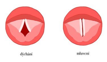
The glottis is widened by the posterior crico-arytenoid muscle. As the only muscle of the larynx, it opens the glottis and keeps it open for breathing - respiratory position . The opposite is phonation position , in which the slit is in a different state. The glottis is narrowed by the crico-arytaenoideus lateralis muscle. The glottis is closed by the arytaenoideus muscle. High-quality closure of the vocal fold is completed by the thyro-arytaenoideus muscle. The vocal cords are shortened by the thyro-arytaenoideus muscle - it is most involved in voice production. The more muscle fibers of the thyro-arytenoid muscle contract, the stronger the vibrating part of the vocal cord is and vice versa. It is active in this mechanismm. vocalis , which is the medial part of m. thyro-arytaenoideus. The vocal cords are stretched and stretched by the crico-thyroideus muscle.
Closure of the lower respiratory tract during swallowing is ensured by three mechanisms:
- extension of the larynx cranially (thanks to the suprahyoid muscles ),
- narrowing of the entrance of the larynx (folding of the epiglottis due to the muscles ending on the epiglottis – ary-epiglotticus and thyro-epiglotticus muscles),
- narrowing of the glottis (due to the crico-arytaenoideus lateralis muscle).
Mucous membrane of the larynx[edit | edit source]
The mucous membrane of the larynx is mostly covered by multi-rowed cylindrical epithelium with cilia . A regular exception is the plicae vocales and part of the surface of the epiglottis , where the epithelium is multi-layered squamous non-keratinized . After birth, the plicae vestibulares are covered with multi-rowed cylindrical epithelium with cilia, but relatively soon islands of stratified squamous non-horny epithelium can appear in them. The islets can be relatively large, and even the plicae vestibulares can be covered by stratified squamous keratinizing epithelium on the histological specimen. The extent of stratified squamous non-keratinizing epithelium can be particularly large in smokers.
In the mucous membrane there are islands of lymphatic tissue and in places even typical lymph nodes. The mucous membrane also contains small seromucinous glandulae laryngeales , but these are absent on the vocal cords.
Vessels and nerves of the larynx[edit | edit source]
Arteries, veins and Lymph[edit | edit source]
- a. laryngea superior (from a. thyroidea superior)
 Arteries of Larynx. [1]
 - a. laryngea inferior (from a. thyroidea inferior)
- vv. laryngeae opening into vv. thyroideae
 Veins of Larynx[2]
 - the lymph drains into the nodi lymphatici cervicales profundi superiores et inferiores
Nerves[edit | edit source]
Motor innervation – comes from the branches of the vagus nerve . It is rich in both motor and proprioceptive components.
- R. externus n. laryngei superioris – innervates one ventral muscle of the crico-thyroideus m.
- N. laryngeus recurrentens – innervates all other muscles.
Sensitive innervation
- R. internus n. laryngei superioris − mucous membrane up to the level of the plica vocales.
- N. laryngeus recurrentens – area under the plica vocales.
- Between the two nerves, there is a so-called Galen's anastomosis on the back surface of the cartilago cricoid.
Parasympathetic innervation – from the vagus nerve.

Sympathetic innervation – from pars cervicalis trunci symphatici.
Links[edit | edit source]
Virtual Microscope[edit | edit source]
Related Articles[edit | edit source]
- Coniotomy
- Tracheotomy
- N. vagus
- Infrahyoid muscles
External links[edit | edit source]
References[edit | edit source]
- PETROVICKÝ, Pavel, et al. Anatomy with topography and clinical applications: Organs and vessels. 1st edition. Osveta, 2001. 463 pp. Vol. 1. ISBN 80-8063-046-1 .
- CIHÁK, Radomír and Miloš GRIM. Anatomy. 2nd 3rd edition. Prague: Grada, 2013. ISBN 9788024747880 .


