Epithelial Tissue

Epithelial tissue consists of closely-packed cells with strong connections to each other and the ECM.
Classification of epithelia[edit | edit source]
Epithelia can be classified in two ways, according to cell arrangement or function:
- cell arrangement
- sheet (membranous, see below for more)
- trabecular
- reticular
- function
- covering
- secretory
- absorptive
- respiratory
- sensory
- myoepithelial
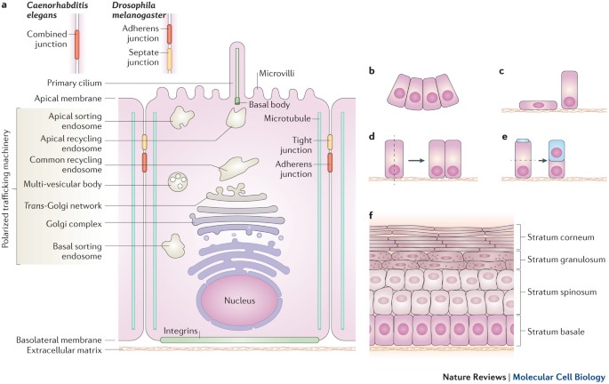
An idealized scheme of epithelial cell with surface domains and modifications appears like this.
For a description of cell surfaces and their characteristics, see Cytology#Cell surfaces and intercellular junctions
Covering epithelium[edit | edit source]
The following are the common types of covering epithelia and their locations in the body:
Simple
- Simple squamous
- endothelium
- mesothelium (serous lining of cavities, pericardium, peritoneum)
- Simple cuboidal
- small ducts of glands
- respiratory bronchioles
- kidney tubules
- covers ovary
- Simple columnar
- large ducts of glands
- GIT
- gallbladder
- oviduct and uterus
- Pseudostratified columnar (all cells attached to basement membrane, nuclei at irregular levels, not all cells reach surface)
- respiratory passages (nasopharynx, larynx, trachea, large intrapulmonary bronchi) and Eustachian tube

Stratified
- Stratified squamous
- keratinized
- epidermis, as well as hard palate and gingiva
- non-keratinized
- mucosa of oral cavity, oropharynx, esophagus, vagina
- keratinized
- Stratified cuboidal
- developing ovarian follicles and sweat glands
- Stratified columnar
- conjunctiva (fornix), male urethra (pars spongiosa)
- Transitional (umbrella cell covers multiple cells below)
- renal calyx, ureter, bladder
Trabecular epithelium[edit | edit source]
- cells are arranged in cords, which in turn form a 3-D network
- capillaries and nerves are arranged amidst the cords
- found in the liver parenchyma, adenohypophysis, suprarenal cortex, islets of Langerhans
- schematic(example from liver)
Reticular epithelium[edit | edit source]
- cells are arranged in a loose mesh work of cells connecting via processes
- nuclei of cells are arranged in the midst of the processes
- found in the thymus and myoepithelium
- schematic (cTEC example from thymus)
Glandular epithelium[edit | edit source]
Glandular epithelium functions in the synthesis and export of various compounds. It can be classified by structure or function.
Structural classification
- Unicellular
- Multicellular
- simple (acinar or tubular)
- branched (also acinar or tubular)
- compound (acinar, tubular, and tubuloacinar)
Functional classification
- exocrine vs endocrine
- merocrine, apocrine, or holocrine
- serous, mucous, or seromucous
Unicellular glands[edit | edit source]
Unicellular glands are modified secretory cells within covering epithelium epithelial tissue of a different kind.

Goblet cells
- Secretes lubricating mucus that aids function of nearby organs
- Major component of mucus is mucin (a glycoprotein)
- Located within covering epithelium of respiratory passages, intestinal mucosa and conjunctiva
Cells within Diffuse Neuroendocrine System (DNES)
- secrete granules particular to cell
- include enterochromaffin cells, argentaffine cells and others
Paneth Cells
- secrete large, eosinophilic granules (lysozyme, α-defensins) into apical cytoplasm
- located within crypts of Lieberkühn of large intestine
Multicellular glands[edit | edit source]
Graphic schematization of the structure and classification of multicellular glands here.
Structures and examples of each morphological type of multicellular glands are given below:
| Structure | Examples | |
|---|---|---|
| Simple | acinar | |
| tubular | Mucous glands of colon, crypts of Liberkuhn | |
| Branched | acinar | Meibomian glands |
| tubular | pyloric and uterine glands | |
| Compound | acinar | lacrimal and parotid glands |
| tubular | duodenal and bulbourethral glands | |
| tubuloacinar | sublingual and submandibular glands | |
Endocrine vs exocrine glands[edit | edit source]
Endocrine glands do not have ducts and secrete hormones into the bloodstream.
Exocrine glands have ducts lined with epithelium that secrete products directly to particular sites. Their different secretion methods are illustrated below.
- Eccrine secretion
- exocytose products
- ex: sweat glands
- Apocrine
- release products (usually lipid droplets) as membrane-enclosed vesicles from cytoplasm
- ex: mammary glands
- Holocrine
- disintegrate secretory cell
- ex. sebaceous glands

Endocrine and exocrine glands also finish development differently.
Serous, mucous, and seromucous glands[edit | edit source]
An overview of the serous, mucous, and seromucous glands is given below:

| Type of Gland | Structure | Cellular properties | Glands |
|---|---|---|---|
| Mucous | tubules | Polyhedral shape. Mucus (carbohydrate) doesn't stain | pyloric and duodenal |
| Serous | acini | Pyramidal shape. Serous proteins are basophilic | lacrimal, parotid, and pancreas |
| Mixed | mucous tubules, serous acini, and serous demilunes (crescents of Giannuzzi) | submandibular and sublingual | |
Absorptive epithelium[1][edit | edit source]
Absorptive epithelium forms the lining of the large and small intestine and the proximal renal tubules.
Cell structure[edit | edit source]
- microvilli form brush border and increase surface area
- tight junctions prevent leaking
- invaginations of basolateral membrane increase surface area and enclose mitochondria
- glycocalyx and brush border slow down flow in the lumen, allowing more efficient use of digestive enzymes
Function[edit | edit source]
- Secretes mucus and gastric juice
- Transports nutrients inward via all types of membrane transport
- Vesicles formed in pinocytosis can fuse with lysosomes or secrete contents at the basolateral surface, where active transport further exports molecules
Respiratory epithelium[edit | edit source]
Conducting zone epithelium
- Pseudostratified columnar ciliated epithelium
- Basal cells renew epithelium
- Goblet cells secrete mucus to moisten tract and protect against mechanical damage and pathogens
- Mucus moved upward via cilia (mucociliary escalator), starting at bronchioles
Respiratory zone (alveolar) epithelium
- Type I (simple squamous) cells function in direct gas exchange
- Type II (cuboidal) cells are interspersed between type I and secrete surfactant
Sensory epithelium[edit | edit source]
There are two types of sensory epithelium:
Primary
- derived directly from neurons
- directly convert stimulus to action potential, signal conducted to CNS
- Ex: olfactory epithelium, rods and cones in retina
Secondary
- External signal interacts with receptors, which then interact with neurotransmitters
- Adjacent neurons send signal to CNS
- Ex: taste buds, vestibulo-cochlear apparatus
Metaplasias[edit | edit source]
Renewal of epithelium is a normal process that constantly happens in the body. Stem cells differentiate into specialized epithelial cells.
Metaplasia is when this renewal happens abnormally - normal epithelium is replaced by a different type. This tissue is unstable, precancerous. Examples include
- Respiratory epithelium
- pseudostratified ciliated --> stratified squamous epithelium
- Can be due to smoking
- Cervical epithelium
- simple columnar --> stratified squamous
- Can be due to viral infections
Dysplasia is when the cells acquire abnormal morphology. This is the first step in cancerous transformation.
Hypertrophy: growth in cell size.
Hyperplasia: growth in cell number.
Neoplasia: regulatory mechanisms are uncoupled, cancerous cells invade normal tissue
References[edit | edit source]
Mescher, A. and Junqueira, L., 2018. Junqueira's basic histology. New York: McGraw-Hill, pp. 71-96.
Vaňhara, Petr et al. Guide To General Histology And Microscopic Anatomy. 1st ed., Masaryk University Press, 2020, pp. 2-3.