Inflammation of the eyelids

Inflammations of the eyelids include hordeolum, chalazion, meibomianitis, inflammations of the lid margins, abscesses and phlegmons, viral and allergic inflammations of the eyelids.
Hordeolum (barley grain)
Hordeolum' (barley grain) is an acute purulent inflammation of the cilia follicles, Moll's or Zeis's gland. The most common causative agents of inflammation are staphylococci'. The first symptoms are "itching" and a feeling of "pressure" in the eyelid. This is followed by "swelling and redness" and the inflammation progresses with the presence of inflammatory infiltration to a purulent "abscess". Subjectively, it is manifested by sensitivity and pain when blinking. Within a few days, the abscess may burst outward.
- Therapy
- Warm tiles, possibly small incisions to facilitate the evacuation of pus.
- Local application of antibiotics or sulfonamides, which prevent the spread of infection.
- Differential diagnosis
Chalazion (wolf grain)
Chalazion is an acute purulent inflammation Meibomian gland located in the area of the tarsal plate. Symptoms of inflammation are more pronounced than in barley grain - we observe redness, soreness and swelling with enlarged preauricular and submandibular nodes. The inflammation can progress to the formation of an abscess or phlegmon of the lid, or it turns into a chronic form.
Chronic chalazion (meibomian cyst) is caused by a blockage of the mouth of the meibomian gland with accumulated secretion. The cyst is painless to palpation, we can see swelling in the tarsal plate of the eyelid. When the upper lid is everted, we see the conjunctiva arching through which a red-purple granuloma shines through, possibly. secondary infection.
- Therapy
- We do not need to intervene if it does not cause cosmetic problems and it heals spontaneously.
- Injection of triamcinolone (0.1–0.2 ml) in combination with a local anesthetic into the swollen tissue.
- If the chalazion does not heal, we repeat the application after 2-3 weeks.
- Generally administered antibiotics for recurrences and especially in patients with acne rosacea or seborrhoeic dermatitis, in whom this disease is more common.
- Unsuccessful conservative therapy - under local anesthesia, an incision can be made perpendicular to the course of the lid and the contents of the chalazion excochleated.
- Differential diagnosis
A Meibomian gland tumor (rare) must be ruled out; if suspected, a biopsy with histology must be performed.
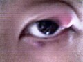
Chalazion of the upper lid

Bleeding chalazion of the lower eyelid

_02.jpg)
Meibomianitis
</noinclude> Meibomianitis is a disease caused by passive retention of Meibomian gland secretions, which occurs in middle-aged population. The first symptom is usually a whitish foamy secretion on the edges of the lids. When massaging the eyelids, we squeeze the sebaceous contents out of the glands. After eversion of the lid, we observe yellowish, vertically placed strips under the tarsal conjunctiva. Deposits of calcium may be present, which pose a danger of Meibomian gland infarctions. We remove them by massaging the lid in the tarsal spot.
Blepharitis (inflammation of the lid edges)

Blepharitis is a chronic inflammation of the edges of the eyelids. The transition of the skin to the mucous membrane of the conjunctiva is very sensitive to various factors. The most common symptom of inflammation is hyperemia, due to the significant vascular supply. Blepharitis is most often caused by staphylococcal infection or it occurs during sebborhoic dermatitis.
Squamous blepharitis
In this form of blepharitis, we observe the formation of scales between the eyelashes and increased blood flow in the entire edge of the eyelids. It takes place as a chronic inflammation accompanied by itching, burning and eye fatigue. Seborrheic eczema often appears on the skin of the eyelids. The cause is often uncorrected or poorly corrected refractive error. We observe squamous blepharitis more often in diabetics, in patients with chronic kidney disease or with inflammation of the alimentary canal. Aggravation of hyperemia manifests itself in smoky, cold or, on the contrary, warm premises.
- Therapy
- Correction of refractive error.
- Removal of irritating factors.
- Mechanical removal of scales with a cotton brush.
- Rubbing the edges of the eyelids with a 3% AgNO3 solution.
- Massage with corticosteroid ointment in combination with antibiotics − sulfonamides.
Ulcerative blepharitis
Ulcerative blepharitis is caused by purulent bacteria, in particular streptococci and S. aureus. In the terrain already affected by squamous blepharitis, purulent deposits begin to form. Hyperemia, redness and swelling of the edges of the eyelids are visible, and we can see dried secretions between the eyelashes. Eyelashes often fall out or scars form on the edge of the lids causing the eyelashes to grow against the bulb. As a result of inflammation, scarred ectropion, epiphora, chronic conjunctivitis occur.
- Therapy
- Local application of antibiotics (sulfonamides).
- In case of severe inflammation, general antibiotic treatment.
Abscesses and phlegmons of the lids
The main causes tend to be:
- Cosmetic eyebrow hair removal;
- progression of acute chalazion;
- infected hematoma;
- infection of small wounds;
- transition of infection from secondary nasal cavities.
The skin in the area of the abscess is tense, strongly reddened, the lid is swollen and warm. The lid is painful and stiff to palpation (it will soften in a few days). The eye slit narrows or is completely closed by swelling. We palpate enlarged preauricular and submandibular nodes. In the progression of the disease, bulb protrusion associated with increased body temperature and even sepsis may occur. The condition can be further complicated by thrombosis of the cavernous sinus.
- Therapy
- Warm compresses (they speed up the size of the abscess);
- antibiotics locally/generally;
- evacuation of the abscess by incision, drainage.
Viral eyelid inflammation
The etiological agent of inflammation in the area of the eyelids is the Herpes simplex virus and the Herpes zoster virus'.
Herpes simplex palpebrae (herpes febrilis)
It begins with edema of the skin of the eyelids, soon a typical vesiculopapular efflorescence is formed. It is caused by the herpes simplex virus. The inflammation is usually one-sided, often after a cold, when the body is generally weakened, or during stress. It heals spontaneously. In case of recurrences, it is advisable to start therapy with acyclovir.
Herpes zooster ophthalmicus (shingles of the first trigeminal branch)
It is a viral infection of the n. trigeminus, Gassersky ganglion or ganglion ciliare that appears unilaterally. The first symptoms are a headache and the appearance of small blisters in the respective innervated area, which burst and turn into crusts. Scars and nerve disorders (hypesthesia, paresthesia) often remain in the places of the original blisters. Even after the skin symptoms have subsided, it is necessary to monitor the condition of the eye due to the possibility of corneal damage caused by keratitis. If the ramus nasociliaris is affected, vesicles also form on the tip of the nose and iridocyclitis, which poses a risk of developing secondary glaucoma.
- Therapy
- Acyclovir.
- Broad-spectrum antibiotics to prevent secondary infection.
- Vitamins B and C.
- Locally indifferent sprinkles, liquid powder, gentian violet.
- Analgesics as needed.
Allergic inflammation of the eyelids - eczema
Eczema arises in connection with the use of cosmetic products, as a reaction to pollen or topical antibiotics. The reaction is most often manifested by itching, redness, swelling.
- Weeping eczema – vesicles are seen that turn into oozing areas. In the chronic form, we observe thickening and thickening of the skin.
- Hyperkeratotic eczema - dry, hardened skin is seen. It very often occurs as contact eczema.
- Therapy (necessary cooperation with a dermatologist)
- Avoiding the trigger.
- Poultices with Jarisch solution.
- Topical steroids.
- Overall antihistamines.
Links
References
- ROZSÍVAL, Pavel, et al. Ophthalmology. 1. edition. Prague : Galén, 2006. 373 pp. ISBN 80-7262-404-0.