Fibula


| HE | A02.5.07.001 |
|---|---|
| Articulating bones | tibia, talus (fibula is not part of the knee joint) |
| Type of articulation | distal end of the fibula and tibia - syndesmosis |
| Function | Muscle spacing |
| Osifikace | chondrogenní |
The fibula (fibula bone) is located laterally compared to the tibia. It serves only as a bone for the beginnings of muscles. We can divide it into four parts:
- caput fibulae – head;
- collum fibulae – neck;
- corpus fibulae – body;
- malleolus lateralis – external ankle.
Head
- Apex capitis fibulae – projection on the head.
- Facies articularis capitis fibulae – contact surface for the tibia.
Neck
The neck is the narrower part between the head and the body.
Body
Like the body of the tibia, the body has a triangular shape. Three faces (facies medialis, lateralis et posterior) and three edges (margo anterior, posterior et interosseus) are distinguished. On the posterior surface of the fibula is the crista medialis – the place between the beginnings of m. ''tibialis posterior and '' ''m. flexor hallucis longus.''
External ankle
It's a little more distal than the medial ankle. On the outer ankle we describe:
- facies articularis malleoli lateralis – contact surface for the ankle bone;
- sulcus malleolaris – groove on the back of the ankle;
- lateral malleoli fossa – fossa for attachment of the talofibular ligamentum posterius.
-
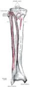 Fibula (left)
 -
 Distal part of the fibula
 -
 Fibula fracture


From the 7th week of pregnancy, ossification begins in the diaphysis (as with the tibia). In the 1st year the distal epiphysis begins to ossify and in the 3rd year the proximal one. A smaller ossification nucleus is also formed in tuberositas tibiae. Between the ages of 15 and 19, both epiphyses grow together with the diaphysis. In girls earlier.
Links
Related articles
Bibliography
- ČIHÁK, Radomír. Anatomie. 2. edition. Praha : Grada Publishing, a.s., 2008. 516 pp. vol. 1. ISBN 80-7169-970-5.

