Tibia


The tibia (tibia) is located medially in front of the calf bone. It has a supporting function. We can divide it into three parts:
- proximal part – condyles (medialis et lateralis);
- corpus tibiae – body bone;
- Distal part – juts out in the malleolus medialis (inner ankle).
Condyles:[edit | edit source]
- The medial condyle is more oval and deeper.
- The lateral condyle is rounder and shallower.
Between the condyles there is the eminentia intercondylaris, in front of and behind the eminence there are places for attachment of cruciate ligaments: area intercondylaris anterior and area intercondylaris posterior.
Other formations on condyles:[edit | edit source]
- Facies articularis fibularis – area for connection with the fibula.
- Tuberositas tibiae – roughness on the anterior surface of the tibia and at the same time the site of the insertion of m. quadriceps femoris (ligamentum patellae). It is the point of contact with the mat while kneeling.
Body[edit | edit source]
The body has a triangular shape. Three faces (facies medialis, lateralis et posterior) and three edges (margo anterior, interosseus et margo medialis) are distinguished. On the posterior surface of the tibia there is a linea musculi solei – a place for the insertion of m. soleus.
Distal part[edit | edit source]
On the distal part we distinguish:
- malleolus medialis (inner ankle);
- sulcus malleolaris – groove behind the inner ankle;
- incisura fibularis – notch for connection with fibula;
- facies articularis – articular surface for connection with the bone ankle;
- facies articularis malleoli medialis – articular surface on the inside of the inner ankle.
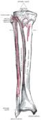
Front side of the tibia

The back of the tibia

Spiral fracture of the tibia

Ossification[edit | edit source]
From the 7th week of pregnancy, ossification in the diaphysis begins. Before birth, the proximal epiphysis begins to ossify, and in the first year the distal diaphysis. A smaller ossification nucleus is also formed in tuberositas tibiae. Between the ages of 15 and 18, both epiphyses and the diaphysis grow together.
Links[edit | edit source]
Related articles[edit | edit source]
Bibliography[edit | edit source]
- ČIHÁK, Radomír. Anatomie. 2. edition. Praha : Grada Publishing, a.s., 2008. 516 pp. vol. 1. ISBN 80-7169-970-5.