Development of the respiratory system
At the beginning of the 4th week, during the development of the respiratory system, the laryngotracheal notch is the first to appear from the ventral side of the foregut. An increased level of retinoic acid (RA - retinoic acid) in the mesoderm induces the expression of the transcription factor TBX4 in the adjacent endoderm, which conditions placement of the laryngotracheal tube. It follows that the epithelium of the respiratory system is of endoderm origin. The remainder (ie, ligament, cartilage and muscle) arises from the mesenchyme of the splanchnopleura which hugs the foregut.
At the beginning, the scrotum communicates widely with the foregut. During its further growth, two longitudinal tracheoesophageal cilia are formed here, which later fuse together to form the tracheoesophageal septum. This separates the esophagus and trachea with the bronchopulmonary lobes.
Larynx[edit | edit source]
Larynx arises from the initial section of the laryngotracheal notch. It is lined with epithelium of entoderm origin. Cartilage and muscles originate from the mesenchyme of the 4th and 6th gill arches. The origin of the muscles explains the innervation of the larynx by means of the vagus nerve (n. laryngeus superior – 4th branchial arch, n. laryngeus recurrentens – 6th branchial arch).
Proliferation of epithelial cells also changes the shape of the entrance to the larynx. An opening in the shape of the letter T arises from the original sagittal slit. During the differentiation of the thyroid and annular cartilages, their shape changes into the aditus laryngis. At the time of differentiation, epithelial cells rapidly proliferate and the opening temporarily closes. The passage is reopened by vacuolation of the epithelium and the bases of the ventriculus laryngis delimited by the plica vocalis and plica vestibularis are formed along the edges.
Trachea, bronchi, lungs[edit | edit source]
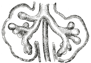
Two bronchopulmonary lobes, which originate from the laryngotracheal lobe, appear at the beginning of the 5. weeks enlarge and form the base of both main bronchi. On the right, the hilum divides into three parts (future secondary bronchi) and on the left into two. Further division into future tertiary bronchi (also segmental) is the basis for bronchopulmonary segments. At the end of the 6th month, approximately 17 divisions are formed, another 6 divisions will take place postnatally. The bronchial tree therefore has 23 divisions.
The lungs grow into the pericardioperitoneal canals. These are finally separated by pleuroperitoneal and pleuropericardial folds from the peritoneal and pericardial cavities. As the lungs grow into the cavity, the mesoderm splanchnopleura gives rise to the visceral pleura and the mesoderm somatopleura becomes the parietal pleura. Since the lungs move caudally during growth, the bifurcation of the trachea lies at Th4 at birth.
Lung Differentiation[edit | edit source]
Lung differentiation can be divided into 4 stages:
- pseudoglandular stage - 5th to 16th week, the bronchial tree branches up to the terminal bronchioles. Respiratory bronchioles and alveoli are not yet formed.
- Canalicular stage - 16th to 26th week, the terminal bronchioles are further divided into two or more respiratory bronchioles and these further into three to six ductus alveolares. Cubic cells lining the bronchioles begin to flatten and alveolar epithelial cells – type I pneumocytes – are formed, to which blood and lymphatic capillaries approach.
- Stage of terminal sacs - 26th week to birth, the formation of terminal sacs (also primitive alveoli). Capillaries are already closely adjacent to the walls of the pouches.
- Alveolar stage - 8th month of fetal development until childhood, mature alveoli are already formed, they are in close contact with capillaries.
Pneumocytes II. type producing surfactant begin to differentiate at the end of the 6th month and the number of capillaries sufficient for respiration is reached during the 7th month. Before birth, the alveoli are filled with a mixture of mucus from the bronchial glands, a fluid with a high chloride content and little protein and surfactant.
Surfactant starts to increase here mainly 14 days before birth. The phospholipids that make it up get into the amniotic fluid, stimulate the macrophages present, and they penetrate the placenta into the uterus, where they produce interleukin 1beta. This substance induces the production of prostaglandins, which stimulate the onset of uterine contractions. Through this mechanism, the fetus affects the beginning of labor.
The fetus performs respiratory movements even before birth. Because of them, amniotic fluid is aspired, they have a great influence on the development of the lungs, and the respiratory muscles are thus also preparing for their future activity.
Lung development continues even postnatally. A newborn's lungs contain around twenty million alveoli, while an adult's lungs contain around 300 million.
Links[edit | edit source]
Related Articles[edit | edit source]
- Congenital developmental defects of the respiratory system
- Development of body cavities, mesenteries and diaphragm
- Trachea
- Lungs
- Larynx
References[edit | edit source]
- SADLER, Thomas W. Langmanova lékařská embryologie. 1. edition. Praha : Grada, 2011. 432 pp. ISBN 978-80-247-2640-3.
- MALÍNSKÝ, Jiří – LICHNOVSKÝ, Václav. Přehled embryologie člověka v obrazech. 2. edition. Olomouc : Univerzita Palackého v Olomouci, 2001. 176 pp. ISBN 80-244-0243-2.


