Development of the cardiovascular system
The vascular system begins to develop during the third week of embryonic development. Blood islands appear first, followed by primitive blood vessels, which merge into primitive blood circulation. The lymphatic system is formed during the fifth week.
Development of primitive blood circulation[edit | edit source]
The development of blood circulation takes place in two stages:
- vasculogenesis
- angiogenesis
Vasculogenesis[edit | edit source]
The first event that leads to the formation of the circulatory system is vasculogenesis. It forms blood islands and the foundations of future blood vessels.
This set of events is controlled by the growth factors, FGF2 (fibroblast growth factor 2), which acts on extraembryonic mesoderm cells and later on intraembryonic mesoderm cells. The action of FGF2 leads to the differentiation and formation of hemangioblasts , which form clusters called blood islands.
Under the influence of another growth factor – VEGF (vascular endothelial growth factor) – differentiation of individual blood islet cells is carried out. This factor is produced by the surrounding mesenchymal cells. VEGF binds to two types of receptors, the type I receptor and the type II receptor, binding first to the type II receptor. This interaction leads to the differentiation of surface hemangioblasts of the blood islet into primitive endothelial cells, the so-called angioblasts. It also acts on cells inside the blood islets that are not in contact with the surrounding mesenchyme, and these turn into the first stem cells called blood stem cells.
VEGF also binds to the type I receptor, leading to the linking of angioblasts and the formation of cellular junctions, primarily zonulae occludentes and this creates the first blood vessels. Inside of this primitive blood vessels primitive blood cells flow, which form hemoglobin but have a preserved nucleus. They therefore remain in the form of erythroblasts.
Angiogenesis[edit | edit source]
The second step leading to the emergence of primitive blood circulation is angiogenesis, which is a set of events that lead to the interconnection of primitive blood vessels and the formation of a blood stream.
This process is again controlled by growth factors, VEGF, TGF-β (transforming growth factor beta), PDGF (platelet growth factor). Under the influence of these factors, primitive vessels grow, new branches sprout and whole networks are formed.
Afterwards a connection of blood vessels arises in several places at the same time :
- in the splanchnopleuric mesenchyme of the extraembryonic mesoderm of the yolk sac
- in the somatopleuric mesenchyme of the extraembryonic mesoderm of the chorion and the germinal shaft
- in the so-called cardiogenic area inside the embryo (in the area of the future heart)
At the end of the third week, all the individual blood vessels are connected and primitive blood circulation is formed .
Development of the heart[edit | edit source]
We can divide the development of the heart into several stages:
- cor tubulare duplex
- cor tubulare simplex
- cor sigmoideum
- embryonic heart
- fetal heart
Cardiogenic area[edit | edit source]
The heart develops in the so-called cardiogenic area , which is the area close to the head of the three-layer structure in the splanchnopleura of the intraembryonic mesoderm. This area is horseshoe-shaped and extends from the primitive knot to in front of the oropharyngeal membrane, where the two sides join to form a complete horseshoe. The formation of this zone is controlled by a combination of different factors.
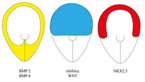
The transcription factor NKX2.5 is expressed in this region . This factor is essential for the formation of heart muscle. Vessels can also form in other areas, but the formation of the myocardial sheath of primitive vessels can only occur in the area of the cardiogenic zone at the site of NKX2.5 expression.
The region with the expression of the transcription factor NKX2.5 is defined by the expression of other factors:
- By expressing BMP 2 and 4 (bone morphogenetic protein), which are produced peripherally in the region of the lateral mesoderm and endoderm .
- By inhibiting the WNT factor, which is normally expressed throughout the trilayer structure, but cardiogenic area its expression is inhibited, due to the action of Crescent and Cerberus inhibitors.
When we overlay the area where BMP 2 and 4 are found and the area where WNT is inhibited, we get a horseshoe shape and thus the area where NKX2.5 is expressed.
Cor tubulare duplex[edit | edit source]
In the area of the cardiogenic zone, blood islands appear during the third week of development, and about one to two days after the formation of these structures in the extraembryonic mesoderm. These blood islands begin to surround the primitive myoblasts. In the course of angiogenesis, a vascular bed is formed. The individual parts begin to fuse and form two endocardial tubes surrounded by a layer of myocardium.
Cor tubulare simplex[edit | edit source]
As the embryo grows, both endocardial tubes begin to move from the cephalic region to the thoracic region. Due to craniocaudal bending, the two endocardial tubes converge until they merge in the midline, giving rise to a unified heart tube . This tube is lined with the endocardium and enveloped by the myocardial mantle , the secretory activity of which creates a temporary layer of heart (cardiac jelly), which separates the endocardium from the myocardium and gives rise to the conduction system of the heart. The bases of the first arteries emerge cranially into the heart tube and the bases of the first veins enter caudally.
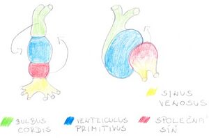
Over time, individual constrictions begin to appear on the heart tube, dividing the entire tube into several parts:
- In the most caudal part, the area of the sinus venosus arises , which is the first compartment of the heart tube as it is initially bifurcated.
- The second part is the heart wall .
- The third section is the area of the ventriculus primitivus , or the primitive chamber, which is separated from the primitive atrium by a narrowing (sulcus atrioventricularis).
- The last part of the heart tube is the bulbus cordis , which is separated from the primitive ventricle by the sulcus bulboventricularis.
This heart tube is already capable of contractions, which appear from the 22nd day. At first they are irregular and move blood in different directions, but by the end of the fourth week they synchronize, allowing blood to flow in one direction.
Cor sigmoideum[edit | edit source]
Cor sigmoideum, or heart loop, is formed by bending the heart tube. This process is completed on the 28th day of development. The heart tube rotates in several directions:
- the region of the bulbus cordis rotates to the right and downward, so that it performs a ventral and caudal movement
- the region of the heart atrium and sinus venosus rotates to the left and up and reaches dorsally behind the bulbus cordis, thus making a dorsal and cranial movement.
This creates the basis for the final shape of the heart.
Embryonic heart[edit | edit source]
It arises from the heart ring due to septation and separation of all definitive parts of the heart.
Fetal heart[edit | edit source]
After the completion of septation, the embryonic heart turns into a fetal heart. This has two separate chambers, the outflow compartment of the heart and the inflow compartment. A small opening, the foramen ovale , remains in the atrial septum, thanks to which the communication between the two atria remains. This is important for the specific blood circulation of the fetus .
Links[edit | edit source]
Related articles[edit | edit source]
- Fetal circulation
- Embryology
- Angiogenesis a neovascularization
- Development of the heart
- Development of the arteries


