Chromosome staining
Chromosome staining is a very important step for and identification. It helps us to classify chromosomes and to describe changes in the chromsome structure.
Classic staining (solid staining)[edit | edit source]
For classic staining, we only use Giemsa Romanovski solution (or Wright's staining). The result of this staining is evenly stained chromosomes without any banding. Thus, we can only classify the chromosomes into groups A, B, C, D, E, F, G. This staining is used nowadays to determine the structural deviations of the chromosomes in people who suffer from congenital chromosomal instability.
G-banding[edit | edit source]
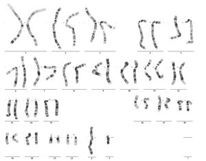
Normal male karyotype, G-banding Using this method, we can not only divide chromosomes into groups, but also distinguish them from each other and determine them precisely. G-banding is based on the action of trypsin and subsequent staining with Giemsa solution. Light and dark bands are now distinguishable on the chromosome. Each G-band has its own number, which is used to describe structural changes and also to locate genes and other DNA sequences . See the picture showing the karyotype of a man.
Other coloring techniques[edit | edit source]
We also know other techniques that are used.
- Q-banding – Here we use the fluorescent dye quinacrine, so we use the abbreviation Q (as in Quinacrine). We get similar results to G-striping, but the method is more expensive. We need a fluorescence microscope to view chromosomes stained in this way.
- R-banding (R-banding, reverse banding) – We use high temperature to partially degrade chromosomes. Then we stain with Giemsa. We get a slightly different result, the R-bands have the opposite color to the G-bands.
- C-banding – We use barium hydroxide and then stain with Giemsa solution. In this way, we can visualize regions of constitutive heterochromatin.
- Staining with colloidal silver (Ag-NOR) – If we want to highlight the p-arms of acrocentric chromosomes, we choose this method. We can therefore investigate aberrations on acrocentric chromosomes (mainly identify so-called markers of acrocentric origin). We use silver nitrate, which acts on proteins located precisely in the neighborhood of NORs (Nucleolar Organizer Regions).
Evaluation of results[edit | edit source]
Computer technology allows us to visualize stained chromosomes. We transfer the image from the microscope to the computer in digitized form. Subsequently, we can sort the individual chromosomes and compile a karyotype.
Links[edit | edit source]
[edit | edit source]
- Identification of chromosomes
- Indications for chromosomal examination
- Hematoxylin-Eosin staining
- Burri staining
- Gram stain
References[edit | edit source]
- KOČÁREK, Eduard – PÁNEK, Martin – NOVOTNÁ, Drahuše. Klinická cytogenetika I. 2. edition. Karolinum, 2010. 134 pp. ISBN 978-80-246-1880-7.

