Body hair and hair
Body hairs and hairs are fibrous keratinized structures found all over the body except the palma manus, planta pedis, red lips, labia minora, clitoris, and glans penis.
There are approximately 60 hairs per cm2 on the body surface . Hairs are the longest body hairs on the human body, they grow on the hairy part of the head in an amount of about 600 hairs per cm2. The character of the hair (color and thickness) is different depending on location, race and can change according to age and gender.
- Lanugo (fluff) – fetal and terminal (hairs), vellus hair, terminal hair;
- pilli longi – mane hair, beard, armpit hair, pubic hair and other pubic hair;
- pilli breves – eyebrows, hairs in the nose, hairs at the mouth of the external auditory canal.
Macroscopic structure[edit | edit source]
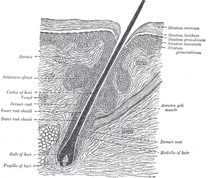
The body hair has two main parts, a free part (scapus pilli) and a part immersed in the skin, i.e. root (radix pilli). In depth, it ends with a thickening, the so-called hair bulb - bulbus pilli with germinal matrix. A part of the fibrous sheath - the hair papilla, which contains numerous blood vessels - sinks into the hair bulb from below.
Microscopic structure[edit | edit source]
The cells of the hair bulb surrounding the hair papilla are cylindrical, sit on a well-developed basal lamina and are equivalent to the cells of the stratum basale epidermis – they divide mitotically and their daughter cells reach the higher regions of the hair itself and its inner epithelial sheath. Another part is the medulla pilli (medulla pilli), which consists of large vacuolated, incompletely keratinized cells. These cells are located in the central part of the bulbus pilli above the hair papilla.
The last part is the hair cortex (cortex pilli), which is made up of cells closely surrounding the central area of the bulbus pilli. These differentiate into spindle-shaped cells, keratinize and remain firmly connected. The layer of cubic cells towards the periphery of the bulbus pilli gradually changes to cylindrical cells, in the direction of the vertical axis they become flattened and completely keratinized. They are placed over each other with their ends, so that their free ends point apically towards the end of the free hair, which resembles the placement of "roof bags".
Melanocytes[edit | edit source]
Melanocytes are dendritic cells of neural crest origin located between epithelial cells at the base of the bulbus pilli. These cells make melanin, which is synthesized in melanosomes from tyrosine and occurs in two forms - eumelanin (the most common form, a brown-black polymer) and pheomelanin (causes red hair and freckles). Hair color depends on the activity of melanocytes. The pigment is transferred to the cells of the cortex and the pith of the hair - in the same way as the keratinocytes in the epidermis.
Hair follicle[edit | edit source]
It is formed by a root, which is surrounded by an outer and inner epithelial sheath. The outer epithelial sheath is a layer of cells from the surrounding skin that sinks into the dermis around the root of the hair and its inner epithelial sheath.
The inner epithelial sheath differentiates from the edge of the bulbus pilli and completely envelops the initial section of the radix pilli. Its cells gradually degenerate and desquamate (they disappear at the level where the sebaceous gland opens into the hair follicle). The inner epithelial sheath consists of three layers:
- Cuticle of the sheath – a similar structure to the cuticle of the hair, but the free ends point in the opposite direction, i.e. towards the bulbus pilli; the cells of both cuticles are wedged together and together they are moved in the apical direction.
- Huxley's layer – 1–3 rows of flattened cuboidal cells that have prominent eosinophilic trichohyaline granules in the cytoplasm.
- Henley's layer - flat epithelial cells that have the character of stratum lucidum epidermis.
In the close area of the apical region of the follicle, there is a fully developed epidermis, which thins out in the deeper parts, and here we find layers corresponding to the stratum germinativum epidermis. Epidermal cells are large bright and rich in glycogen. Around the outer epithelial sheath we find a fibrous sheath composed of inner circularly and outer longitudinally arranged elements of fibrous tissue, capillaries and nerve fibers are located between the two layers.
After the hair falls out, a new bulbus pilli is formed at the end of the cords of cells of the outer epithelial sheath, a newly formed fibrous papilla with blood vessels grows into this structure, and the growth of a new hair begins.
Musculus arrector pilli[edit | edit source]
It is a bundle of smooth muscle cells that bridges the sebaceous gland. It is attached to the fibrous sheath and to the stratum papillare corii and straightens the hair with its contraction. In addition, it facilitates the emptying of the sebaceous gland and causes depression of the skin where it is embedded in the dermis ("goosebumps").
Links[edit | edit source]
Related arcticles[edit | edit source]
- Anatomy of the skin • Physiology of the skin
- Skin adnexa • Nails
- Sebaceous glands • Apocrine glands • Ecrinne sweat glands
- Histology: Thick-type skin (histological preparation) • Axilla/histological preparation
Source[edit | edit source]
- BENEŠ, Jiří. Studijní materiály [online]. ©2007. [cit. 30.11.2010]. <http://jirben2.chytrak.cz/>.
References[edit | edit source]
- ŠTORK, Jiří. Dermatovenerologie. 1. edition. 2008. ISBN 978-80-7262-371-6.


