Types of sutures in surgery
From WikiLectures

This article has been translated from WikiSkripta; ready for the editor's review.
Split Stitches[edit | edit source]

Single stitch

Single stitch

Mattress stitches

Matress stitches

Mattress stitches

Subcutaneous suture


In terms of time[edit | edit source]
- Primary suture
- immediate – wounds clean , uninfected, with smooth edges, suture immediately after wound treatment.
- deferred - wounds suspected of infection, suture 3 to 7 days after excision before granulations are formed, if there is no infection in the wound.
- Secondary suture
- early - wounds dirty and infected, suture after 2 weeks , if the edges are cleanly granulated, mobile, when the infection has been controlled, the edges do not excise.
- postponed - dirty and infected wounds that have suppurated for a long time, suture after 3 weeks after excision of immovable granulated edges to loosen them and "bleed".
In terms of surgical technique [ edit | edit source ][edit | edit source]
- Single (single-knot) stitch and its modifications
- mattress stitch horizontal (U-stitch) and vertical (Donatti stitch);
- Algower suture (partially intradermal vertical mattress suture);
- intradermal;
- astringent (tobacco - more correctly snuff box);
- Out of stitches.
- Continuation stitch
- not transferred (modification - intradermal, ...);
- tossed around.
Needles[edit | edit source]
- Bent needle – various parts of the circular section, most often:
- semicircular (1/2 circle) – for suturing in depth (under the skin), organs (anastomoses on the GIT, vessels, ...);
- semi-oval (3/8 circle) – cutting needle for sewing leather;
- J-shaped – sutures in depth, e.g. suturing the abdominal wall in the opening after the laparoscopic port.
- Straight needle – e.g. basic stitch for tendon suture.
- We also distinguish between classic needles (with an eye into which the thread is threaded) and atraumatic (the thread is sealed at the end of the needle and it does not cause excessive damage when it penetrates the tissues).
- The cross-section of the needle can be :
- triangular (cutting needles) – suture of the skin , subcutaneous tissue, fascia and muscles;
- round - mucous membrane, parenchymal organs and digestive tubes, blood vessels;
- round with a triangular tip (so-called tapercut) – for fascia, ligaments.

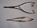
Mayo-Hegar needle
Microsurgical needles

Atraumatic needle


Threads[edit | edit source]
Thread properties[edit | edit source]
- Tensile strength - depends not only on the strength of the fiber, but also on its configuration and material, two scales are used to indicate strength :
- European (EP) – indicates the fiber strength in tenths of mm (e.g. EP 1 means a fiber with a thickness of 0.1 mm);
- American (USP designation) - strength is indicated by the number of zeros (the more zeros, the weaker the fiber), the number of zeros is usually indicated by the number before the slash followed by a zero - i.e. the lower the number before the slash, the stronger the fiber - e.g. 3/0 and 4/0 are used to suture the skin on the trunk and limbs, 5/0 on the face, 8/0 – 10/0 fibers to suture vessels and nerves.
- Thread configuration
- monofilament fiber – penetrates the tissue smoothly, less tendency to colonize microbes, elastic – the disadvantage is that the knot is easily untied, greater memory – after the end of the fiber is cut, it sticks out and irritates the surroundings;
- polyfil fiber (braided or woven from several fibers) – easier to handle, the knots hold better, they do not loosen and the ends do not stick out after the fiber is cut (suitable for skin eyelashes), the disadvantage is capillarity, which allows infection to penetrate into the wound (on the other hand, this fiber can also be used as a capillary drain) and also scrubbing during tissue penetration (this can be reduced by coating with a synthetic material - e.g. coated Vicryl).
- Fiber memory – the ability to return to its original shape after deformation.
Fiber material[edit | edit source]
- Unabsorbable
- silk (Mersilk) – polyfilic, easily tied, does not cut, to skin sutures and ligatures;
- polyamide = nylon, silon (Ethilon, Peterlone) – monofil and polyfil, special type – oriented silon (orsilon);
- polypropylene (Prolene) – for vascular reconstructions, absolutely non-absorbable;
- polyester = dacron (Mersilene) – also absolutely non-absorbable (100% strength indefinitely);
- steel wire - bone surgery, coated when securing laparotomy (Ventrofil).
- Absorbable
- natural (enzymatic decomposition - an inflammatory reaction occurs around the fiber) - catgut (purified collagen ) - according to EU standards, it can only be used for veterinary purposes;
- synthetic (hydrolytic decomposition - no inflammatory reaction):
- polyglycolic acid ;
- polyglactin 910 (Vicryl) ;
- polydioxanone (PDS II) .
- Metal clips and clamps - most often during laparoscopy (treatment of blood vessels, bile ducts, appendix stump ), suturing the skin with metal clips, loading silver clamps on brain aneurysms .
Principles for suturing individual organs[edit | edit source]
- the use of fibers of different materials, different strengths and configurations in different locations and situations is dependent on the practices of the given workplace, operator and material equipment, hereafter only general principles:
Skin suture[edit | edit source]
- Atraumatic fibers are used for suturing the skin (i.e. sealed to the end of the needle, which thus smoothly continues directly into the fiber), a so-called skin needle (3/8 circle, with a tip of a triangular cross-section - cutting) is used.
- Metal clamps can also be used.
- Types of stitches:
- single-node – simple, mattress, intradermal, Algower sutures, …
- ongoing – mostly for intradermal sutures.
- Material:
- non-absorbable – for all sutures knotted above the surface of the skin (including continued intradermal ones);
- absorbable - only for individual intradermal sutures.
- A good adaptation of the edges of the wound is necessary, this can be achieved by gently pulling on the edges of the wound or the nearest sutures, using adaptation tweezers (like surgical ones, but with more teeth).
- Stitch removal depends on location:
- face - in 3 to 4 days;
- trunk, limbs - in 7 to 10 days;
- above the joints (generally in places of great tension) - in 10 to 14 days.
Subcutaneous suture[edit | edit source]
- Necessary for incisions in areas with a lot of subcutaneous fat (it is of fundamental importance for the appearance of the resulting skin scar) - it is performed with an absorbable material on a semicircular cutting needle , the aim is to bring the edges of the skin closer together and prevent the formation of pockets in the subcutaneous tissue, where the hematoma could accumulate and later become infected , in the event of a large leakage of blood, it is necessary to drain it under the skin (miniredon taken out of the wound, capillary drain taken out directly through the wound - it is removed in 24-48 hours).
Fascia and muscle suture[edit | edit source]
- When suturing a muscle, the fascia plays a major role in the strength of the suture - we don't sew the muscle at all or only with a few adaptation stitches.
- A semi-circular cutting needle, absorbable and non-absorbable material, longitudinally cut fascia is sufficient to be sutured with a continuation suture (prevention of muscle hernia), longitudinal incision through the muscle (e.g. when closing an oblique incision after appendectomy ) – the suture can ischemia the muscle, it is sufficient to close it with several individual sutures, knotted not under tension and perpendicular to the course of the muscle fibers, transverse section (cut through the muscle belly) – individual sutures of the fascia, or place several mattress sutures around the perimeter of the muscle and then bring the edges of the muscle closer together with longitudinal individual sutures.
- When closing the hernia gate, we sew the fascia with knotted sutures with a strong non-absorbable material , it is possible to use a roof plastic according to Mayo (extensive umbilical hernias ), it is possible to sew in two layers, in case of strong tension and in recurrent hernias, the so-called tension- free suture using a mesh is used .
Tendon suture[edit | edit source]
- Monofilament absorbable or non-absorbable fibers, or fine wire.
- It is necessary to create a smooth surface of the tendon so that adhesions do not occur (also early rehabilitation):
- suture of flexors - modified suture according to Kirchmayr ;
- extensor suture – similar to the flexor suture, but usually a mattress U-stitch is sufficient .
Vascular suture[edit | edit source]
- Monofilament atraumatic thread (Prolene) thin up to 10/0 equipped at both ends with an atraumatic needle, or PTFE fiber, material absorbable by the growing organism.
- The thread is perfectly smooth, it is necessary to tie at least 6 knots and leave longer ends.
- It is possible to sew with individual stitches (especially with a growing organism - a continuous stitch is too inflexible), the most used is a continuous non-overlapping stitch, the sewn ends of the vessels must lie exactly on each other (clamped vessels are sewn, blood flow is restored after suturing and we monitor whether blood leaks through the anastomosis ), the suture covers all layers of the vessel wall (vessels are dissected subadventitally), knots are generally made from the outside.
- Because of the risk of thrombosis, heparin (1 to 2 mg/kg) is administered in total before the vessel is clamped , after the reconstruction is completed it is neutralized with protamine .
- Ligatures:
- absorbable fibers (smaller vessels, mainly veins - saphenous vein ligation during varicose vein surgery, etc.);
- non-absorbable fibers (larger vessels).
Suture of nerves[edit | edit source]
- Very thin monofilament non-absorbable fiber, or tissue adhesives (autologous plasma) can be used.
- The suture is performed with individual sutures behind the epineurium in immediate operations, behind the perineurium in delayed operations (the epineurium must be cut so that it does not get stuck between the cutting surfaces).
- It is important to precisely fit the cutting surfaces (the perineurium of the peripheral end of the nerve represents the guiding structures through which axons grow), we must not allow mutual rotation of both ends of the nerve, the sutured ends must not be bruised or otherwise damaged (in this case, we cut them with a razor scalpel).
- In the case of defects over 2 cm, an autograft must be used (most often the suralis nerve).
- The nerve grows at a rate of approx. 1 mm/day, it takes 4-6 weeks to grow through the sutured area (in the case of autotransplants, a delay of 8-12 weeks), the denervated muscle undergoes irreversible atrophy after 1 year.
Suture on the digestive system[edit | edit source]
- End-to-end, side-to-side, or end-to-side alimentary canal anastomoses raise two issues that are still debated:
- single or continuous suture?
- suture in one or two layers?
- The best answer is to use a suture that the surgeon knows and trusts.
- So-called intestinal needles (semi-round with a round tip) and absorbable fibers (preferably monofilament or coated knitted) are used.
- Among the series of stitches bearing the names of their (sometimes real) authors, the following are known and used:
- Albert's suture - occupies all layers of the alimentary canal, knotted externally;
- Lembert suture (seromuscular) – involves only the serosa and muscularis externa;
- Mikulicz's stitch - occupies all layers, knotted inside.
- Various combinations are possible (for double-layer sutures, e.g. Albert-Lembert suture).
- The main complications of GIT sutures are:
- stenosis of the alimentary canal;
- suture insufficiency (fistula formation).
- The modern way is suturing using staplers - linear or circular, their advantage is the speed of the suture, the disadvantage is the high price (e.g. the removal of hemorrhoids according to Long is not covered by the insurance company, unlike the classic operation according to Langenbeck), the main indication for stapler suture is the need to quickly treat an intestinal injury within polytrauma.
- Another modern option for end-to-end bowel connection is the use of an absorbable ring (Valtrac).
Suture of parenchymal and other organs[edit | edit source]
- Fragile parenchymal organs - the danger of cutting through the sutures - the sutures can be underlaid or several mattress sutures can be placed along the edges of the wound areas parallel to them, and only then the wound can be sutured with additional sutures perpendicular to the previous ones.
- Needles with a round cross-section, with the largest possible diameter, are used.
- Bladder suture - absorbable fiber in two layers, must not pass into the lumen of the bladder (formation of concretions).
Wound closure after laparotomy[edit | edit source]
- The possibility to sew in anatomical layers or take several layers at once with one stitch:
- peritoneum – continuous tossed suture with absorbable material;
- fascia and muscles – for the strength of the suture, the most important thing is the suture of the fascia ( rectus abdominis vagina in middle laparotomies, aponeurosis of the external oblique muscle in alternating incisions), the abdominal muscles are either not sutured at all or are approached with several stitches perpendicular to the direction of the fibers;
- under the skin – individual stitches with absorbable material;
- skin - non-absorbable suture (single knot, continuous, intradermal).
- A newer method is the loop suture from PDS II - a needle with a double thread, a continuous suture that involves the peritoneum, the posterior sheath of the vagina, the muscle, the anterior sheath of the rectus abdominis muscle and part of the subcutaneous tissue - this suture is mechanically the strongest and, moreover, separate the suture of the individual layers still ends with a common scar.
Links[edit | edit source]
Sources[edit | edit source]
- PASTOR, Jan. Langenbeck's medical web page [online]. [cit. 7. 12. 2009]. <https://langenbeck.webs.com/>.