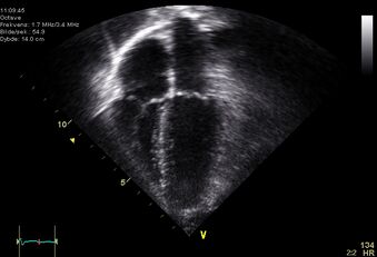
Two-dimensional echocardiography
From WikiLectures
It shows flat "cuts" of the heart while maintaining the actual movement of individual structures.
Indication:
- Demonstration of pathological cardiac structures– thrombi, myxoma, tumors;
- assessment of valve structure and movement - post-rheumatic and congenital valve defects, mitral valve prolapse, forward movement of the anterior mitralis in hypertrophic obstructive cardiomyopathy, infectious endocarditis;
- evaluation of ventricular systolic function by assessment of myocardial systolic thickening - coronary heart disease, cardiomyopathy, heart defects, myocarditis;
- myocardial infarction complications - rupture of the tendons / walls of the left ventricle, aneurysm;
- morphological + structural abnormalities - congenital heart defects;
- pericardial effusion and tamponade
- pericardial diseases - constrictive pericarditis;
- heart wall thickness - hypertension.
Links[edit | edit source]
Related articles[edit | edit source]
- echocardiography
- stress examination of the cardiovascular system
References[edit | edit source]
CHILD, P., et al. Internal Medicine. 2nd edition. Prague: Galén, 2007. ISBN 978-80-7262-496-6.

