Thyroid
The thyroid gland (lat. glandula thyreoidea ) is a butterfly-shaped endocrine organ located on the front of the neck. It is functionally involved in the regulation of metabolism by producing the hormones trijodtyroninu a thyroxine. These hormones influence metabolic rate, oxygen consumption, growth and development. Another hormone produced by the thyroid gland is calcitonin, which regulates the metabolism of calcium and phosphorus.
Anatomy[edit | edit source]
The gland consists of two lobes ( lobus dexter et sinister ) in the shape of a triangular pyramid connected by an isthmus. Variably , in 1/4 of the cases there is a lobus pyramidalis - a remnant of the ductus thyroglossus departing cranially from the isthmus .
- Lobes: 5–8 cm long, 2–4 cm wide, 1.5–2.5 cm thick.
Weight is usually 20–60 g (larger in females).
- There are differences in the size and weight of the thyroid gland according to gender, age, but also geographic differences (on average, the gland increases with increasing distance from the sea and with rising altitude).
The surface is smooth to slightly bumpy, has a reddish-brown to reddish-purple color (depending on the blood vessels). It consists of a fibrous sheath containing two leaves.
- Superficial list: capsula externa (related to the fibrous sheath of the cervical neurovascular bundle and lamina praetrachealis fasciae colli).
- Deep sheet: the capsula propria sends a septa to the gland, which divides it into lobules (lobules). They further consist of pouches, follicles separated from each other by fine tissue and capillary and lymphatic meshes.
- Between the two leaves are vascular braids.
The shape of the gland is variable. Depending on the location of the isthmus, it can have the shape of the letter H, U, or A. The base of the individual lobes is usually caudally rounded, the tips point cranially.
Syntopy of individual anatomical surfaces of the lobes[edit | edit source]
The gland is located on the sides of the larynx a trachea, the isthmus lies in front of the 2nd to 4th tracheal rings. It is fixed to the trachea with ligamentum suspensorium glandulae thyroideae s. ligamentum Berry.
Syntopy of individual anatomical surfaces of the lobes[edit | edit source]
Facies medialis: attached to the larynx and trachea, relation to nervus laryngeus recurrens.
Facies anterolateralis : covered together with the isthmus by infrahyoid muscles from the lamina pretrachealis fasciae colli.
Facies dorsalis : relation to the cervical neurovascular bundle, partially embedded in it are the parathyroid glands.
Vascular supply[edit | edit source]
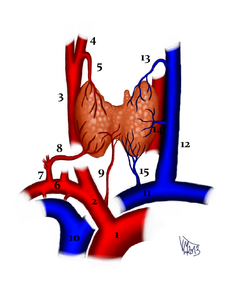
The cranial pole of the lobes is supplied by the superior thyroid artery (5), a branch from the external carotid artery (4), the caudal pole by the inferior thyroid artery (8), a branch from the thyreocervicalis truncus (7). Both arteries form a delicate anastomosis.
![]() An inconstant a. thyroidea ima (9) from the truncus brachocephalicus (2) or from the arch of the aorta (1) may be present.
An inconstant a. thyroidea ima (9) from the truncus brachocephalicus (2) or from the arch of the aorta (1) may be present.
The veins are collected in a plexus between the capsula externa and the capsule propria. Blood leaves the upper pole via vv. thyroideae superiores (13) and vv. thyroideae mediae (14) to internal jugular vein (12). From the lower pole, the blood drains vv. thyroideae inferiores (15) to v. brachiocephalica sinistra (11).
Lymphatic vessels lead to the subcapsulation of the plexus, and subsequently to the nodi lymphatici cervicales profundi.
Microscopic structure[edit | edit source]
Microscopically, the gland is made up of spherical, closed follicles . The wall of the follicles consists of one layer of cubic follicular cells, which can become flattened with hypofunction of the gland, while hyperfunction is manifested by a change in the shape of the cells to a cylindrical shape. The follicles are richly interwoven with sinusoids. All follicular cells sit on the basement membrane, their cytoplasm is basophilic, rich in rough endoplasmic reticulum and secretory granules.
Follicles are filled with colloid . It is a viscous, homogeneous liquid containing especially the glycoprotein thyroglobulin, which conditions the positive PAS reaction of the colloid. Thyroglobulin contains precursors of the main thyroid hormones - thyroxine and triiodothyronine, whose synthesis process takes place in the colloid.
In addition to follicular cells, parafollicular cells (C-cells) producing calcitonin .are also found in the thyroid gland . We can find them between follicular cells either individually or in groups of 2-3 cells. Their cytoplasm contains large amounts of HER and electron-dense granules. Developmentally, they classically originate from the endoderm of the 4th gill slit, not from the neural crest, as previously reported.
Links[edit | edit source]
Related Articles[edit | edit source]
- Examination of the function of the thyroid gland
- Thyroid disease
- Examination of the thyroid gland diseases
- Thyroid hormones
References[edit | edit source]
- PETROVICKÝ, Pavel. Anatomie s topografií a klinickými aplikacemi. 1. edition. Martin : Osveta, 2001. 560 pp. 2; ISBN 80-8063-047-X.
- JUNQUIERA, L. Carlos – CARNEIRO, José – KELLEY, Robert O.. Základy histologie. 1. edition. Jinočany : H & H 1997, 1997. 502 pp. ISBN 80-85787-37-7.


