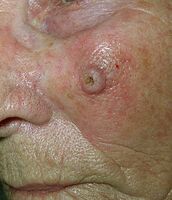
Spinalioma
Squamous cell carcinoma is also one of the malignant epithelial tumors of the skin. It usually begins with intraepithelial growth, followed by destructive progression . It metastasizes mainly through the lymphatic system. The incidence in our population corresponds to about 11 / 100,000, compared to basal cell carcinoma it occurs less frequently (approximately 1:10).
Etiology[edit | edit source]
It usually develops from precancerous lesions (solar keratosis, leukoplakia, Bowen's disease, etc.), especially in predisposed individuals, with a lower amount of melanin in the skin (phototype I and II). Other risk factors are chronic degenerative skin changes ( scars , fistulas , skin ulcers ), immune disorders (immunosuppression), HPV infections and long-term exposure of the skin to carcinogens .
Clinical Picture[edit | edit source]
The clinical picture of the tumor develops over time.
- form: Diffuse infiltrating (inconspicuously elevated hyperkeratosis or solid, infiltrated deposit with a bumpy surface, grows slowly, metastasizes late);
- form: Ulcerative (in the center there may be disintegration and ulceration with swollen, stiff edges);
- form: Exophytic (soft, aggressively and rapidly growing formation with central decay and bleeding, early metastasis).
Metastases to regional nodes occur in about 5-10% of patients. The nodules tend to be rigid, large in size, with the possibility of ulceration and fistula formation.
We recognize special forms with their typical precancerous lesions:
- Lip augmentation (from leukoplakia and cheilitis);
- vulvar spinalioma (from lichen sclerosus et atrophicus );
- penile spinalioma (from erythroplasia );
- tongue spinalioma (from leukoplakia and erythroplakia );
- aggressively growing exophytic form (from Bowen 's disease and chronic irritation).
Diagnostics[edit | edit source]
Histopathological examination is the decisive examination for determining the diagnosis . Tumor prognosis correlates with the degree of cell dedifferentiation.
Therapy and prognosis[edit | edit source]
Radical excision with the edge of healthy tissue (to the periphery and depth). For smaller lesions, an intact edge about 1 cm wide is recommended. The excision of larger tumors should have a margin of healthy tissue 2-3 cm, especially on the trunk, where spinalioma behaves more aggressively and the solution of the resulting defect does not cause major difficulties. In the case of metastases to regional nodes, we also perform their removal.
If surgery is impossible (eg in the elderly), we perform radiotherapy. In the presence of metastases, we supplement with chemotherapy.
The prognosis depends on the location, size of the tumor and the degree of dedifferentiation. Tumors in the solar area have the best prognosis, worse on the ear, lips and scars. Tumors located on the mucous membranes have the worst prognosis.
Links[edit | edit source]
Related Articles[edit | edit source]
- Malignant skin tumors : Melanoma Basal cell carcinoma Verrucous carcinoma
- Benign skin tumors
- Precancerous lesions in dermatology
- Malignant mesenchymal tumors: Kaposi's sarcoma Dermatofibrosarcoma protuberans
Resources[edit | edit source]
- JANEČEK, VLADIMÍR. SPINALIOM [online]. [feeling. 2011-02-09]. < http://www.liposukce.cz/plasticka-chirurgie/kozni-nadory/spinaliom.htm >.
- ŠTORK, Jiří, et al. Dermatovenerology. 2nd edition. Prague: Galén, 2013. 502 pp. ISBN 978-80-7262-898-8 .


