Spinal medulla
__CONTENTS__ The spinal cord forms the center for simple reflexes. It runs through the spinal canal at the level of C1–L2 (length 40–50 cm) and is covered by spinal sheaths. It fulfills reflex and conversion functions''. The anterior and posterior spinal roots emerge from it, which then merge into a nerve bundle. The spinal cord contains mixed fibers' (motor and sensitive) and vegetative fibers.
Spinal cord anatomy[edit | edit source]
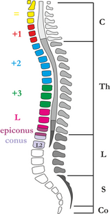
At the height C2–T2 there is thickening – intumescentia cervicalis and at the height T12–L1 sub> intumescentia lumbosacralis (lumbalis).
The spinal cord is divided into spinal segments. A spinal segment is a section of the spinal cord from which 1 pair of spinal nerves converges (a total of 31 pairs of spinal nerves – 8 cervical, 12 thoracic, 5 lumbar, 5 sacral, 1 coccygeal). Caudally, it narrows conically into the 'conus medullaris', the tip of which extends to L1–L2 then continues as a bundle of nerves, which we call ' 'cauda equina.
Structure of spinal cord[edit | edit source]
- White matter
-
- Funiculus lateralis, anterior and posterior – cords in which nerve fibers run upwards and downwards.
- Fissura mediana anterior – anterior notch between the anterior horns.
- Sulcus medianus posterior (posterior notch).
- Sulcus anterolateralis – the fibers of the anterior roots of the spinal cord emerge from it.
- Sulcus posterolateralis – exit of the fibers of the posterior roots of the spinal cord.
- On the back root of the spinal cord there is a node ganglion spinale' (envelops the bodies of sensitive neurons).
- Gray matter
- Arranged in an H shape, formed by a cluster of neurons, it forms the front and back corners of the spinal cord'. The lateral horns of the spinal cord are formed by the columns columnae anteriores, laterales and posteriores (the anterior ones contain motoneurons, the lateral vegetative neurons, the posterior connecting neurons). The spinal canal runs through the center - canalis centralis.
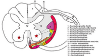
- Spinal cord section.png
Spinal cord section - regions
- Spinal tracts.png
Spinal tracts
Spinal reflexes[edit | edit source]
Spinal reflexes can occur at the level of the spinal cord. The spinal cord ensures emptying of the bladder and rectum. The anatomical basis of the reflex is reflex arc. The spinal cord is subordinate to the brain in its activity, it is the lowest reflex center in terms of development, its interruption means failure of reflexes.
- 5 parts
- receptor, sensor;
- centripetal track (sensitive);
- CNS;
- centrifugal track (motorized);
- effector.
Spinal roots[edit | edit source]
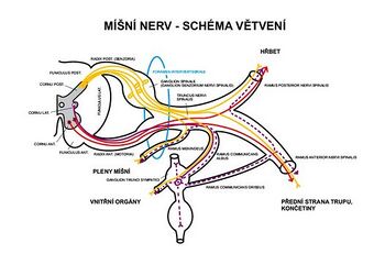
The root fibers, fila radicularia ventralia, come from the sulcus ventrolateralis and connect to the anterior spinal roots - radices anteriores. The "fila radicularia dorsalia" emerge from the dorsal part of the spinal cord and connect to the posterior spinal roots - "radices dorsales". The roots enter the foramen intervertebrale and join the nervus spinalis.
Centrifugal fibers lead to the anterior roots of the spinal cord, centripetal fibers to the posterior roots of the spinal cord. In terms of function, the front spinal roots are motor' and the posterior spinal roots are sensitive.
Spinal nerves[edit | edit source]
- Plexus cervicalis (C1–C4) – sensitively innervates the scalp and supraclavicular region, motorically innervates the muscles of the neck (e.g. n. phrenicus).
- Plexus brachialis (C4–Th1) – innervation of the upper limb (e.g. n. radialis, ' 'n. ulnaris).
- Thoracic nerves - passes inside the intercostal spaces, does not form any plexus. They innervate the chest wall.
- Plexus lumbalis (L1–L5) – innervation of the skin and muscles of the abdomen, thighs and pelvis.
- Plexus sacralis (S1–S5) – innervates the back of the thigh, buttocks, lower leg and leg. The thickest nerve in the human body n. ischiadicus.
Links[edit | edit source]
Related Articles[edit | edit source]
External links[edit | edit source]
- Medulla spinalis (Czech Wikipedia)
- Spinal cord (English Wikipedia)
- Mefanet - Atlas of spinal cord sections
References[edit | edit source]
- CIHÁK, Radomír. ANATOMY 3. Second, revised and supplemented edition. Prague : Grada, 2004. 692 pp. ISBN 80-247-1132-X.


