Optical microscopy (3LF)
- Previous chapter: 6.1 MICROSCOPY
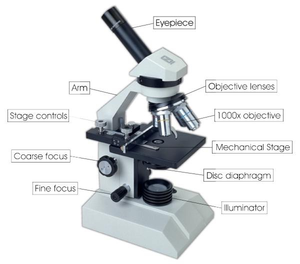
Optical or light microscopy involves passing visible light through or reflected from the sample through a single lense or multiple lenses to allow a magnified view of the sample. The resulting image can be detected directly by the naked eye, imaged / visualized on a photographic plate or captured digitally camera/by a digital camera. In the past there were classical photographic techniques , today are used digital CMOS or CCD cameras for obtaining image.today we use digital CMOS or CCD cameras in order to obtain images
Limitations of standard optical microscopy (bright field microscopy) lie in three areas;
- The technique can only visualize dark or strongly refracting objects effectively.
- Diffraction limits resolution to approximately 0.2 micrometres (see: microscope).
- Out of focus light from points outside of the focal plane reduces image clarity.
Living cells in particular generally lack sufficient contrast in order to be studied successfully, internal structures of the cell are colourless and transparent. The most common way to increase contrast is to stain the different structures with selective dyes, but this often involves killing and fixing the sample. Staining may also introduce artefacts, apparent structural details that are caused by the processing of the specimen and are thus not a legitimate features of the specimen.
In order to get better images there are several sophisticated technologies, that can bring better resulting images. Phase contrast microscopy used for imaging living cells, fluorescence microscopy used for tracing fluorescence markers inside specimen, confocal microscopy and more.
Links[edit | edit source]
Next chapter: 6.1.2 Electron microscopy
Back to Contents

