OZPP module - Practicum No. 1

From WikiLectures

This article was checked by pedagogue
This article was checked by pedagogue  

This article ws checked by pedagogue, but later was changed.

Checked version of the article can be found here.

See also comparation of actual and checked version.

Changed checked article.png


Přehled
Preparation No. 1 - van Gieson staining (elastic fibers).png Preparation No. 2 - van Gieson staining (elastic fibers).png HE (myocardium) 4x.jpg HE (myocardium) HE (myokard) 20x..jpg Lipid staining (liver) prukaz tuku (jatra) 4x.jpg
Z1-10 lipid staining (liver) prukaz tuku (jatra) 20x oznaceno.jpg Grocott (mycosis) Grocott (prukaz plisne) 4x.jpg Grocott (mycosis) Grocott (prukaz plisne) 20x.jpg Z1-3 blue trichrome (kidney) modry trichrom (ledvina) 4x oznaceno.jpg Z1-3 blue trichrome (kidney) modry trichrom (ledvina) 20x oznaceno.jpg
Z1-4 van Gieson (gallbladder) van Gieson (zlucnik)4x oznaceno.jpg Z1-4 van Gieson (gallbladder) van Gieson (zlucnik) 20x oznaceno.jpg Reticulum (liver) retikulum (jatra) 4x.jpg Reticulum (liver) retikulum (jatra) 20x.jpg Z1-7 Mowry (appendix) Mowry (apendix) 4x oznaceno.jpg
Z1-7 Mowry (appendix) Mowry (apendix) 20x oznaceno.jpg PAS (clear cell renal cell carcinoma) PAS (Grawitzuv ca) 4x.jpg Z1-8 PAS (clear cell renal cell carcinoma) PAS (Grawitzuv ca) 20x oznaceno.jpg Z1-9 Pearls reaction (lung) prukaz zeleza (plice) 4x oznaceno.jpg


Preparation No. 1 and No. 2 - van Gieson staining (elastic fibers)

Preparation No. 1 - van Gieson staining (elastic fibers).png Preparation No. 2 - van Gieson staining (elastic fibers).png

Structures

  • tunica intima
  • tunica media
  • tunica adventitia

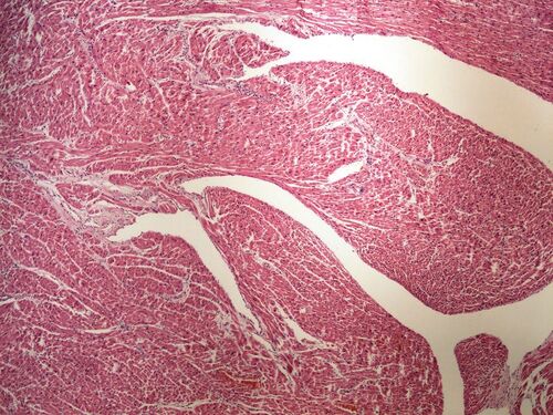
Preparation no. 3 and no. 4 – myocardium (HE)

HE (myocardium) 4x.jpg HE (myocardium) HE (myokard) 20x..jpg

Structures

  • cardiomyocytes
  • box-shaped nuclei of cardiomyocytes

Preparation no. 5 and no. 6 – fat identification (liver)

Lipid staining (liver) prukaz tuku (jatra) 4x.jpg Z1-10 lipid staining (liver) prukaz tuku (jatra) 20x oznaceno.jpg

Structures

  • intracytoplasmic fat vacuoles in hepatocytes

Preparation no. 7 and no. 8 – silvering according to Grocott (proof of mold)

Grocott (mycosis) Grocott (prukaz plisne) 4x.jpg Grocott (mycosis) Grocott (prukaz plisne) 20x.jpg

Preparation no. 9 and no. 10 – blue trichrome staining (kidney)

Z1-3 blue trichrome (kidney) modry trichrom (ledvina) 4x oznaceno.jpg Z1-3 blue trichrome (kidney) modry trichrom (ledvina) 20x oznaceno.jpg

Structures

  • collagen fiber

Preparation no. 11 and no. 12 – van Gieson staining (gallbladder)

Z1-4 van Gieson (gallbladder) van Gieson (zlucnik)4x oznaceno.jpg Z1-4 van Gieson (gallbladder) van Gieson (zlucnik) 20x oznaceno.jpg

Structures

  • smooth muscle
  • ligament

Preparation no. 13 and no. 14 – staining for reticular fibers (liver)

Reticulum (liver) retikulum (jatra) 4x.jpg Reticulum (liver) retikulum (jatra) 20x.jpg

Structures

  • reticular fibers

Preparation no. 15 and no. 16 – staining according to Mowry (appendix vermiformis)

Z1-7 Mowry (appendix) Mowry (apendix) 4x oznaceno.jpg Z1-7 Mowry (appendix) Mowry (apendix) 20x oznaceno.jpg

Structures

  • lymphatic follicle with a germinal center
  • intracytoplasmic mucus vacuoles in mucosal lining cells

Preparation no. 17 and no. 18 – PAS staining (Grawitz's approx.)

PAS (clear cell renal cell carcinoma) PAS (Grawitzuv ca) 4x.jpg Z1-8 PAS (clear cell renal cell carcinoma) PAS (Grawitzuv ca) 20x oznaceno.jpg

Structures

  • PAS positive glycogen granules in the cytoplasm of tumor cells

Preparation no. 19 and no. 20 – detection of iron by Perls reaction (lungs)

Z1-9 Pearls reaction (lung) prukaz zeleza (plice) 4x oznaceno.jpg Z1-9 Pearls reaction (lung) prukaz zeleza (plice) 20x oznaceno.jpg

Structures

  • alveoli
  • positive siderophages in the iron test according to Perls

Links

Navigation

Practicum No. 2 →