Native record and provoking tests
Native Record (NO EVOKED POTENTIALS)
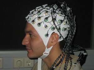
However the term describes the creation of an electroencephalogram using the method of electroencephalography. First observed over 80 years ago it measures and records electrical signals, created by the neurons of the cortex, using small disks of silver chloride as electrodes attached to the scalp. In routine examination 16 unipolar channels are used, with the reference electrode usually attached to the ear.
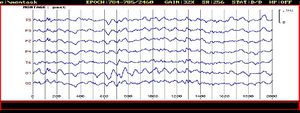
EEG signals are very low, between 5 to 100 µV. Recording them on an electroencephalogram, not only voltage amplitude is to be seen but also the frequencies, which lead to the term brainwaves, both of them dependent on the mental activity of the brain examined. A normal EEG can look like this:
You can distinguish 4 main types of waves:
Beta F: 15-20Hz, A: 5-10 µV (subject in alert state)
Alpha F: 8-13 Hz, A: > 50 µV (subject in relaxed state)
Theta F: 4-7 Hz, A: < 50 µV (subject in deep sleep, children)
Delta F: 0.5-4 Hz, A: 100 µV (pathological)
Why is this method of such a great medical importance? Even though the synchronicity between the actual brain activity and the measured signals on the surface is still of unknown extent, it makes brainmapping(relate certain areas to certain assignments) possible and through this allows the diagnosis of various malfunctions of the cortex, such as epilepsy, brain tumors, parkinson, learning disabilities, migrane and to determine the brain activity (death) during coma. This is partly performed by analyzing the electrical signals that result from the brain receiving external stimuli, evoked potentials, using :
Provoking Tests (EVOKED POTENTIALS)
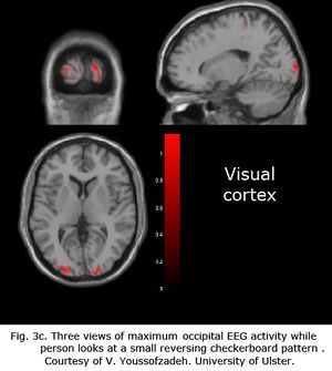
Early visual stimuli to be used for testing evoked potentials were developed in several laboratories in the 1970s, for example by A.M Halliday, who projected a reversing checkerboard pattern onto a translucent screen, still the most common today(with the difference in the use of a video monitor). Recording the response as EEG is made possible by simple computer programs which extract only the visually evoked potential. The stimulations happens in primary visual cortices but also secondary areas, usually recorded by channels from occipital scalp overlying the calcarine fissure.
In analyzing migrane and seizures (epileptic) you subject the examined persons to strobe light during brainwave measurements to look if a change in the EEG can be seen, “activating” the EEG. A commercial stroboscopic stimulator is placed approx 1 m from the patient’s eyes, which are closed and straight looking.The test is performed by alternating flashes varying from 1 to 35 Hz and lasting for 10 s, interrupted by 10 to 30 s with no stimulation. In healthy people a response can be any level of “driving" in the occipital leads. The first response appears shortly after the stimulator starts and stops when the stimulator stops. Also a photomyogenic response is normal, widespread muscle twitching appears, timed to the stimulator. In case of a neurological illness the stimulation would lead to f.e. seizures shortly after the stimulation started. Next to visual, auditory (click or tone presented through earphones) and tactile or somatosensory (tactile or electrical stimulation of a sensory or mixed nerve in the periphery) stimuli are used to provoke potential.
What does the future hold?
While scientists are exploring how non-invasive electrical brain stimulation kits could alleviate the symptoms of Parkinson’s disease or depression, market researchers use EEG for relating neural measurements to choice prediction for future products.
References :
"Biophysical principles of medical technology" - Prof.MUDr. Ivo Hrazdira, DrSc. Doc. RNDr. Vojtech Mornstrein, Csc. MUDr Ales Bourek ; Brno 2000 ; ISBN 80-210-2414-3
http://webvision.med.utah.edu/book/electrophysiology/visually-evoked-potentials/


