Nail diseases
Pathological changes manifesting in the nail unit form a relatively large group of diseases and disease symptoms that can accompany a number of skin and systemic diseases. However, they can also occur independently.
Non-neoplastic dystrophic changes[edit | edit source]
Anonychia[edit | edit source]
Congenital or acquired absence of the nail plate. It accompanies, for example, lichen planus, epidermolysis bullosa, trauma.
Pterygium unguis[edit | edit source]
When the nail plate is lost, the lateral nail ridges grow into the area of the nail bed.
Beau's Lines[edit | edit source]
Transverse grooves in the nail plate as a sign of temporary growth arrest (in case of infection, trauma).
Leukonychia[edit | edit source]
It belongs to a group called chromonychia (changes in the color of the nail bed). Leukonychia punctata et linearis is a commonly occurring chromonychia caused by minor trauma (e.g. manicure).
Mallet fingers[edit | edit source]
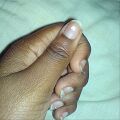
Also called pulmonary hypertrophic osteoartopathy or digiti Hippocratici. They occur on the hands and feet. It accompanies lung diseases, such as emphysema or malignant tumors. They can also be a symptom of heart failure. In club fingers, the so-called Lovibond angle (the angle between the proximal nail ridge and the nail plate, physiologically it is less than 165°) is pathologically enlarged, which can be seen when looking at the nail from the lateral side. We can also confirm the diagnosis with the so-called Schamroth test, placing the same fingers of both hands (e.g. both index fingers) with the distal joints dorsal to each other so that the nail plates touch. Physiologically, the so-called Schamroth window is created as a consequence of the Lovibond angle.
Psoriatic nails[edit | edit source]
We find typical dimpling of the nail plate and so-called oil stains of the nail bed.
Half and half nail[edit | edit source]
The proximal half of the nail bed is light, the distal half is dark red. It indicates renal insufficiency or uremia. It is reversible, disappears with treatment.
Kolionychia[edit | edit source]
Also spoon-shaped nails, the free edge of the nail plate evertes, most often accompanies sideropenic anemia or thyroidopathy. It is also a part of Plummer-Vinson syndrome.
Onychogryphosis[edit | edit source]
Marked deformity with bizarre shapes of the nail plate, bent down or to the sides.
Onychoschisis[edit | edit source]
Lamellar fraying of the free edge of the nail plate.
Infectious diseases[edit | edit source]
- See the Paronychium page for more detailed information
- See the Onychomycosis page for more detailed information
Tumors and cancerous affections of the nails[edit | edit source]
Benign tumors[edit | edit source]
Koenen's tumor[edit | edit source]
It is a periungual fibroma, occurs in patients with tuberous sclerosis.
Digital (acral) fibrokeratoma[edit | edit source]
A hard skin growth growing near the nail unit.
Subungual exostosis[edit | edit source]
A painful bony growth growing from the dorsal side of the distal phalanx (especially the big toe) may elevate the nail plate, often asymmetrically. We demonstrate on X-ray.
Glomus tumor[edit | edit source]
A painful tumor showing through under the nail plate as red blood, occurs especially in women, painful symptoms are manifested mainly in the cold. Glomus tumor cells arise from transformed smooth muscle cells of the so-called Sucquet-Hoyer canal, which is a kind of arteriovenous anastomosis at the fingertips with a thermoregulatory function. Histologically, we distinguish three types of glomus tumors - glomangioma (the most common), solid glomus tumor and glomangiomyoma.
Pyogenic granuloma[edit | edit source]
It is a lobular capillary hemangioma, a necessary biopsy to differentiate it from the amelanotic form of acrolentiginous melanoma.
Onychomatricom[edit | edit source]
Visible as a longitudinal yellow stripe of the nail plate, described relatively recently.
Digital mucoid cyst[edit | edit source]
In fact, a pseudocyst without its own wall, contains a viscous fluid.
Subungual verruca[edit | edit source]
A hyperkeratotic condition caused by HPV infection.
Keratoacanthoma[edit | edit source]
A rapidly growing tumor with typical central hyperkeratinization.
Malignant tumors[edit | edit source]
Squamous cell carcinoma[edit | edit source]
Subungual melanoma[edit | edit source]
It is a type of acrolentiginous melanoma, manifested by so-called melanonychia (a type of chromonychia, a pigmented longitudinal strip of the nail plate). Melanonychia is often associated with pigmentation of the proximal nail fold = Hutchinson's sign
Gallery[edit | edit source]
Links[edit | edit source]
Related articles[edit | edit source]
References[edit | edit source]
RICH, Phoebe A – SCHER, Richard K, et al. An Atlas of Diseases of the Nails. - edition. Parthenon Publishing Group, 2003. 136 pp. ISBN 1-85070-595-X.


