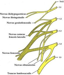
Lumbar plexus
The lumbar plexus is formed by a network of nerve fibers supplying the lower limb. It is located near the Psoas major muscle at the lumbar region.
The plexus is formed by fibers emerging from the anterior rami of vertebral L1-L4 and receives contributions from thoracic vertebra T12.
Branches
The fibers from L1-L4 create cord, which combine to create the 6 major nerves of the Lumbar plexus, which descend down the posterior abdominal wall to reach the lower limb.
The main branches are:
- Iliohypogastric nerve
- Ilioinguinal nerve
- Genitofemoral nerve
- Lateral femoral cutaneous nerve
- Obturator nerve
- Femoral nerve
Iliohypogastric Nerve
The Iliohypogastric nerve is the first major branch arrising from the lumbar plexus. It runs to the iliac crest, crossing the quadratus lumborum muscle, and piercing through the transversus abdominis.
Roots: L1 (with contributions from T12).
Motor Functions: Innervates the internal oblique muscle and transversus abdominis muscle.
Sensory Functions: Innervates the posterolateral gluteal skin in the pubic region.
Ilioinguinal nerve (L1)
The ilioinguinal nerve follows the same general path as the iliohypogastric nerve. It is usually slightly smaller. It continues through the superficial inguinal ring to innervate the skin of the genitalia and middle thigh.
Roots: L1.
Motor Functions: Innervates the internal oblique and transversus abdominis.
Sensory Functions: Innervates the skin on the superior antero-medial thigh. In males, it also supplies the skin over the root of the penis and anterior scrotum. In females, it supplies the skin over mons pubis and labia majora.
Genitofemoral nerve
After exiting the psoas major muscle, the genitofemoral nerve divides into a genital branch, and a femoral branch. It can divide anywhere from inside the Psoas major muscle or only near the Inguinal ligament.
Roots: L1, L2.
Motor Functions: The genital branch innervates the cremasteric muscle.
Sensory Functions: The genital branch innervates the skin of the anterior scrotum (in males) or the skin over mons pubis and labia majora (in females). The femoral branch innervates the skin on the upper anterior thigh.
Lateral femoral cutaneous nerve
This nerve only has sensory (cutaneous) innervation. It enters the thigh at the lateral aspect of the inguinal ligament, where it provides cutaneous innervation to the skin.
Roots: L2, L3
Sensory Functions: Innervates the anterior and lateral thigh down to the level of the knee.
Obturator nerve
After its formation, the obturator nerve descends through the fibres of the psoas major and emerges from its medial border. It then travels posteriorly to the common iliac arteries and laterally along the pelvic wall – towards the obturator foramen of the pelvis.
The obturator nerve enters the medial thigh via the obturator canal (formed between the obturator foramen by the obturator membrane). It then divides into an anterior and posterior division:
- Anterior division: Found anterior to adductor brevis muscle. Descends between the adductor longus and brevis, then pierces the fascia lata, becoming the cutaneous branch of the obturator nerve.
- Posterior division: Found posterior to adductor brevis muscle. Pierces the obturator externus, descending between the adductor magnus and brevis
Roots: L2, L3, L4.
Motor Functions: Innervates the muscles of the medial thigh – the obturator externus, adductor longus, adductor brevis, adductor magnus and gracilis.
Sensory Functions: Innervates the skin over the medial thigh.
Femoral nerve
Nervus femoralis is formed by fibers from the roots of L2−4. It passes through the isthmus at the lacuna musculorum. Vulnerable places of this nerve are during its pelvic course on the lateral side of the psoat (pelvic tumors, laparoscopy), in the lacuna musculorum and in the fossa iliopectinea. It motorically innervates the iliopsoas, sartorius and quadriceps femoris muscles, providing sensitive innervation on the inner side of the thigh and the inner side of the lower leg. Allows for hip flexion and knee extension.
Image of polio
- high nerve damage
- palsy m. iliopsoas (flexion disorder in the hip) and m. quadriceps femoris (extension disorder in the knee) − cannot step, cannot climb stairs
- quadriceps atrophy
- low nerve damage
- extension damage in the knee − the knee breaks (especially when walking down stairs), walking is unstable
- sensitivity disorders in the innervation area (inner thigh and lower leg)
Causes
- pelvic trauma - fractures, dislocations
- consequence of surgery - hip joint surgery, extirpation of inguinal nodes, etc.
- iatrogenic - wrong application of i.m. injections, hematomas after angiography
- pressure in the area of inguinal canal - tumors, enlarged nodes, aneurysm a. femoralis
Links
Related Articles
Link
- PASTOR, Jan. Langenbeck's medical web page [online]. [cit. 2009]. <https://langenbeck.webs.com/>.
- AMBLER, Zdeněk – BEDNAŘÍK, Josef. Klinická neurologie : část speciální. II. 1. edition. Praha : Triton, 2010. ISBN 978-80-7387-389-9.
Links
Related Articles
References
- The Lumbar Plexus - Spinal Nerves - Branches - TeachMeAnatomy. teachmeanatomy.info/lower-limb/nerves/lumbar-plexus/.
- ČIHÁK, Radomír. Anatomie III. 3. edition. Grada, 1997. 672 pp. ISBN 80-7169-140-2.


