Larynx, trachea and bronchi - structure and function
Larynx (larynx)[edit | edit source]
The larynx is the first compartment of the lower respiratory tract and is lined with airway mucosa. The skeleton is composed of a series of cartilages. The large cartilages are hyaline, which ossify in adulthood. The small cartilages are elastic.
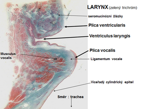
The laryngeal cavity arches in 2 lobes:
- upper lash: plica vocalis
- lower lash: plica vestibularis
Between them runs a fold, the ventriculus laryngis.
The entire mucosa of the larynx, except for the plica vocalis, is covered by a multi-rowed cylindrical epithelium with cilia and goblet cells. The lamina proprii mucosae' of the plica vestibularis is composed of sparse collagenous connective tissue containing collagen fibres and fibroblasts, and also contains small seromucinous glands that often extend to the cartilaginous submucosa.
The vocalis lung is covered by a layered squamous epithelium that does not thicken. Beneath the epithelium on the plica vocalis, glands are absent; there is very sparse connective tissue into which elastic fibers run from the ligamentum vocale (elastic ligament). Laterally, bundles of striated muscle run musculus vocalis.
In the fold of the larynx (ventriculus laryngis), seromucinous glands are abundant in the lamina propria mucosae and there is an accumulation of elements of the lymphatic lineage, sometimes organized into lymphatic follicles (tonsilla laryngea).
Trachea (trachea)[edit | edit source]
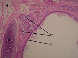
Trachea and bronchi are the continuation of the lower airway. The trachea is a tube reinforced by ring-shaped hyaline cartilages (15-20). The cartilages are open posteriorly, connected by a connective membrane (pars membranacea tracheae) connected by ligaments and circularly arranged bundles of smooth muscle (tunica fibro-musculo-cartilaginea).
The trachea is divided into pars cervicalis, which is located in the anterior part of the visceral space. It further contains dorsally - oesophagus, ventrally - isthmus glandulae thyroideae, infrahyoid muscles, vv. thyroideae inferiores (mouth to plexus thyroideus impar - mouth to v. brachicephalica), laterally - lobi glandulae thyroideae, n. Pars thoracica in the upper mediastinum, also contains dorsally - oesophagus, ventrally - arcus aortae, from which branches to plexus briachiocephalicus (dx.), a. carotis communis sin, a. subclavia sin., thymus. Laterally - v. cava inferior (dx.), v. azygos, n. laryngeus recurrens, n. vagus dx. i sin. The trachea also touches the pulmo dx. separated by the mediastinal pleura. The boundary of the pars cervicalis and pars thoracica is defined by the upper margin of the manubrium sterni, apertura thoracis superior.
The syntopia of the beginning of the trachea is projected at the level of the C6 vertebrae, as a continuation of the larynx (C5 in infants and C4 in neonates) and ends as the bifurcatio tracheae at the level of the Th4-5 vertebrae.
Arterial supply: rr. tracheales (a. thyroidea inferior), rr. bronchales (aorta abdominalis)
Venous supply: vv. tracheales
Lymphatic supply: nodi lymphoidei paratracheales, nodi lymphoidei tracheobronchales
Nerve supply: PS - n. laryngeus recurrens (n. vagus)
S - ggl. stellatum
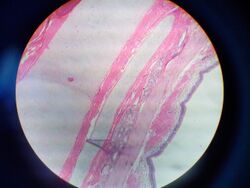
The wall of the trachea is composed of 3 layers. Tunica mucosa - covered by multilayered cylindrical epithelium, except for the bifurcation, which contains mostly layered squamous nonconfluent epithelium. On the mucosal surface there are small seromucinous glands glandulae tracheales' that extend into the submucosa. In the lamina propria mucosae there are islands of lymphoid tissue. In contrast to the alimentary canal, where the muscularis muscularis mucosae forms a boundary, a layer of longitudinal elastic fibres (conus elasticus) separates the lamina propria mucosae from the tunica submucosa. The submucosa attaches either directly to the perichondrium of the hyaline cartilage or to the tunica muscularis. Tunica fibro-musculo-cartilaginea is composed of 15-20 cartilagines tracheales, which are connected by ligg. anularia. The trachea with the cartilago cricroidea is connected by lig. cricotracheale. The posterior side of the trachea is formed by the pars membranacea and bundles of smooth muscle oriented longitudinally and transversally form the m. trachealis. Tunica adventitia continues into the inersticial ligament into the mediastinum. It connects the trachea to the environment and also allows its movements during respiration. It contains sparse collagenous connective tissue.
Bronchi (bronchi)[edit | edit source]
Bronchioles (histology)
Bronchi branch and the cartilage gradually shrinks until only small fragments remain at the bifurcation points. The main supporting component of the wall consists of circularly or spirally arranged bundles of smooth muscle, supplemented by elastic and reticular fibres. The epithelium also gradually decreases from multilayered cylindrical epithelium with cilia to single-layered cylindrical in the terminal bronchiole, which lacks kinocilia, and to cuboidal to squamous epithelium in the respiratory bronchioles. There are neither goblet cells nor glands, but Clara cells appear. The apical part of the cells of the Clara cells is convex and contains secretory granules and a smooth endoplasmic reticulum, which is massively developed here. The Golgi complex, the granular endoplasmic reticulum, which in contrast is not very well developed, and the mitochondria are in the basal part and near the nucleus. These cells produce surfactants (lipoproteins and CC16 protein) that prevent luminal adhesion during expiration. At the sites of bronchial bifurcation, cells of the neuroendocrine system DNES or chemoreceptors are found.
References[edit | edit source]
Related articles[edit | edit source]
Literature[edit | edit source]
- MARTÍNEK, J. – VACEK, Z.. Histologický atlas. 1. edition. Grada, 2008. ISBN 978-80-247-2393-8.
- KONRÁDOVÁ, V. – UHLÍK, J. – VAJNER, L.. Funkční histologie. 2. edition. H&H, 2000. ISBN 80-86022-80-3.
- JUNQUEIRA, L.C.U. – MESCHER, A.L.. Junquiera´s Basic Histology. 12. edition. McGraw-Hill Medical, 2010. ISBN 9780071630207.


