Granuloma
Granulomatous inflammation is a distinctive pattern of chronic inflammation encountered in a limited number of infectious and non infectious conditions. It is a cellular attempt to contain an offending agent which is difficult to eradicate. Typically there is strong activation of T lymphocytes, leading to activated macrophages, resulting in tissue injury.
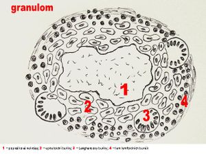
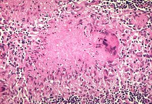
A granuloma is thus a focus of this chronic inflammation consisting of a microscopic aggregation of macrophages that are transformed into epithelial-like cells (called histiocytes, or epithelioid cells), with variable numbers of lymphocytes and plasma cells. The epithelial-like cells (histiocytes) may often fuse to form multi-nucleated giant cells.
Types of Granulomas[edit | edit source]
3 Basic Histological Types Based on Morphology[edit | edit source]
- Foreign body granulomas: will see foreign material within histiocytes/giant cells, sometimes called “foreign body giant cell reaction” by pathologists
- Caseating granulomas: granulomas that induce cell mediated immune response and thus have central necrosis; these are usually associated with infection (e.g. mycobacterial,fungal infections)
- Non-caseating granulomas: granulomas that induce cell mediated immune response without central necrosis (e.g. Sarcoidosis, Crohn’s disease)
Epitheloid Granuloma[edit | edit source]
A sharply circumscribed nodule that mainly consists of epitheloid cells(modified macrophages).
- Tuberculous granuloma.
- Sarcoidal granuloma.
Histiocytic Granuloma[edit | edit source]
An ill-defined nodule that consists of phagocytic histiocytes(tissue macrophages).
- Foreign-body granuloma.
- Rheumatoid granuloma.
- Rheumatic granuloma.
Morphology of Granulomas[edit | edit source]
Sarcoid Granuloma[edit | edit source]
Small granulomas that mostly consists of epitheloid cells. No necrotizing center, but fibrosis may be present. The outer layer consists mostly of collagen. Sarcoid granulomas can occure in:
- Sarcoidosis.
- Crohn`s disease.
- Berylliosis.
- Extrinsic Allergic Alveolitis.
- Primary Billiary Cirrhosis.
Tuberculous Granuloma[edit | edit source]
A large circumscribed granuloma consisting of epitheloid cells around a caseous necrotic core with interspersed Langhans cells. The outer layer consists mostly of lymphocytes. Tuberculous granulomas can occure in:
- Tuberculosis.
- Leprosy.
- Syphilis - A granuloma in syphilis is called gumma. They have coagulative necrosis and central vessels in their core, and plasma cells in their peripheral zone.
Pseudotuberculous Granuloma[edit | edit source]
As the name implies, they are quite similar to tuberculous granulomas. They are ill-defined granulomas consisting of macrophages and epitheloid cells. Granulocytes(mostly neutrophils) are present in the caseous core. They may form abscesses. Pseudotuberculous granulomas can occure in:
- Yersinia Pseudotuberculosis.
- Brucellosis.
- Listeriosis.
- Histoplasmosis.
- Cryptococcosis.
- Typhoid fever.
Rheumatic Granuloma[edit | edit source]
A granuloma with specialized macrophages(called Anitchov cells)around a core of fibrinoid collagen necrosis. Aschoff cells(a variant of giant cells) are interspersed between the other cells, while lymphocytes make up the outer layer. Rheumatic granulomas mostly occure in myocardium and only in rheumatic fever.
Rheumatoid Granuloma[edit | edit source]
A granuloma with a core of fibrinoid collagen necrosis, surrounde by a wall of epitheloid cells. Several centimeters in diameter. Lymphocytes are present in the outer layer. Often occuring in multiple subcutaneous locations and articular nocules in rheumatoid arthritis.
Foreign-Body Granuloma[edit | edit source]
A granuloma with epitheloid cells surrounding a material that cannot be broken down, or that provides large enough difficulties in doing so. The foreign body is surrounded by epitheloid cells and giant cells. The outer layer consists of lymphocytes, fibroblasts and vessels.
Links[edit | edit source]
Related Articles[edit | edit source]
External Links[edit | edit source]
Bibliography[edit | edit source]
- WERNER, Martin – GROSSMANN, John. Color Atlas of Pathology. 1. edition. 2004. 457 pp. ISBN 1-58890-117-3.
- MACFARLANE, Peter S. Pathology Illustrated. 5th edition. 2000. ISBN 9780443059568.


