Genetics of Ig, B and T receptors
The recognition of foreign antigen is made possible by the existence of two types of molecules acting as receptors:
- T cell receptor,
- B cell receptor and derived immunoglobulins.
Immunoglobulins - Ig[edit | edit source]
Glycoproteins occurring as:
- Anchored in the plasma membrane of B-lymphocytes as membrane or surface Ig (mIg) forming receptor,
- freely present in blood, lymph and tissue fluids.
Contact between mIg and foreign antigen is necessary to induce the production of free antibodies. Most B cell receptors are composed of Ig of the IgM and IgD types.
T cell receptors (TCR)[edit | edit source]
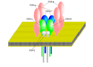
TCR heterodimers are associated with additional polypeptides with which they form a TCR-complex = a group of 3-5 polypeptides that are required for TCR expression on the surface of the T lymphocyte and for signal transduction into the cell interior. Heterodimer composed of 2 polypeptide chains:
- 95% - α and β chains (TCR2),
- 5% - γ and δ chains (TCR1).
TCR2[edit | edit source]
TCR2 α and β chains are transmembrane polypeptides, composed of an outer part that is anchored in the plasma membrane by a transmembrane portion and a short cytoplasmic region.
B receptor genetics[edit | edit source]
The ability of the immune system to recognise antigens depends on receptor molecules on B and T lymphocytes; both cell populations can recognise a huge number of antigens. The cellular and molecular processes leading to this diversity are as follows for both receptor types:
First and foremost, they are changes that have accumulated over the course of evolution and are passed on from generation to generation, where the original gene has been "multiplied" by a process of repeated duplications. At the same time, mutations have taken place that have diversified these genes so that they are not exact copies of the original gene (thus creating the so-called gene family). If these different genes remain in one chromosomal region, they form a gene complex - this contains many genes or segments that code for variable regions and only one or a few segments that code for a constant region. Within the complex, further changes (somatic diversification) take place in somatic cells, but these are not transmitted to the offspring (these are various rearrangements of the genes of the complex in individual cells, resulting in a definitive stretch of DNA encoding a particular receptor).
B receptor genetics[edit | edit source]
Immunoglobulin chains are encoded by three gene complexes:
- IgH - for the heavy chain (chromosome 14),
- IgK and IgL for the light chain κ [kappa] (chromosome 2) and λ [lambda] (chromosome 22).
IgK complex[edit | edit source]
A gene complex consisting of three regions:
- Variable (VK),
- the junction (JK),
- constant (CK).
The constant region is connected by 1 gene, the others are made up of a larger number of genes. Each V gene has 2 exons separated by a short intron. The first exon determines the so-called leading polypeptide sequence, which is required for antibody transport through the endomembrane system of the cell. The second exon encodes the variable part of the antibody.
Each lymphocyte contains more than 100 IgK segments, but only three of these are translated into the language of the protein: one of the VK genes, one of the JK genes and the CK gene, which is made possible by the rearrangement of segments during B cell development. 1 of the VK genes is linked to 1 of the JK genes and a deletion of DNA occurs, which is deposited between the selected genes. These rearranged genes are transcribed into mRNA, including sequences between the VK-JK combination and the CK segment. These sequences are cleaved as introns during post-transcriptional editing. The mature mRNA contains a specific V-J combination linked by a C segment.
Deletion of the 'excess' DNA during several gene rearrangements is made possible by the existence of both V and J. This is referred to as a somatic mutation.
IgL complex[edit | edit source]
A gene complex of a different arrangement. In addition to many Vλ genes, it contains 6 Cλ genes. Each C gene has its own segment J. During rearrangements, any of the V genes combine with any of the J-C pairs. Each lymphocyte carries information for both types of light chains. However, immunoglobulins produced by a single cell have either a κ or λ chain, never both at the same time. This phenomenon is called allelic exclusion.
IgH complex[edit | edit source]
A complex containing at least 100V, 20D and 5J segments in humans. The C region here consists of several segments - Cμ,Cδ,Cγ,Cε,Cα. Depending on which C gene is functional, the resulting immunoglobulin can be classified into classes (Cμ - IgM,Cδ - IgD, etc.).
Conversion steps: fusion of DH with one JH gene. One of the VH genes is subsequently relocated to this junction. After the rearrangement of these segments is completed, transcription of these segments continues towards the C region, the Cμ gene. Transcription stops, resulting in a primary transcript from which all non-coding sequences are excised. After translation, the heavy chains of μ are formed in the cell and associate with the light chains, and the complete IgM molecule is exposed on the cell surface. Some primary transcripts contain both Cμ and Cδ information. The Cμ region is excised and after translation the cell produces the δ heavy chains and the complete IgD molecule. All these processes occur during the development of the lymphocyte from precursors into mature B cells and then stop. After the B lymphocyte encounters antigen, DNA rearrangements continue during proliferation.
Allelic exclusion[edit | edit source]
Each antibody consists of 2 identical heavy and 2 identical light κ or λ chains. Ig molecules produced by a single cell have the same specificity for the antigen and have identical V-regions. Thus, in each B-lymphocyte, the Ig gene complex of only one of chromosomes 2 or 22 (for the light chains) and one of chromosome 14 (for the heavy chains) is active; the other Ig gene complexes of homologous chromosomes are excluded from function - they do not undergo segment rearrangements or transcription = so-called allelic exclusion (one allele of the pair is not active).
Ig variability[edit | edit source]
The enormous variability of antibodies is made possible by three mechanisms:
- if we assume that the V, D and J segments can combine randomly, then 107 different combinations can arise (thus 107 different antigens can be distinguished),
- the possibility of inaccurate joining of segment ends during the joining process; some nucleotides may be deleted or inserted,
- somatic mutations, which occur when the antigen-stimulated B-lymphocyte begins to proliferate; errors may occur during DNA replication that may escape repair mechanisms.
T receptor genetics[edit | edit source]
T receptor je kódován třemi genovými komplexy:
- TCR-α located on chromosome 14,
- TCR-β on chromosome 7,
- TCR-γ on chromosome 7.
The δ-chain genes (TCR- δ) are interspersed between the TCR-α genes.
The TCR-α gene complex is composed of 3 regions: variable (Vα), junctional (Jα) and constant (Cα). The C-region contains 1 gene, the others are composed of a large number of genes. The order of the regions is V-J-C. In addition, there is a D segment with a different arrangement of genes in the TCR-β complex. The D-J-C segment is duplicated here: Vn-D1-J1-C1-D2-J2-C2. Each lymphocyte then contains several hundred TCR segments, but only 7 of them are translated into proteins: 1Vα, 1Jα and 1Cα; 1Vβ, 1Dβ, 1Jβ and 1Cβ. This results in 2 polypeptide chains, α and β, forming a receptor in the plasma membrane.
During T-lymphocyte development, segment rearrangement occurs as follows:
- One of the Vα genes is fused to one of the Jα genes, and a deletion of DNA that is sandwiched between the selected genes occurs. Within one of the duplicated D-J-C segments, one of the Dβ genes is fused to one of the Vβ genes, again resulting in deletion of the DNA segments. The genes thus rearranged are transcribed into mRNA, introns are cleaved out, and editing into mature mRNA is completed; deletion of "extra" DNA during gene rearrangements is made possible by the existence of signal sequences flanking each V, D, and J segment.
The signal sequences are made up of:
- heptamers - palindromic sequences of 7 base pairs: CACAGTG (GTGTCAC),
- spacers - consisting of either 12 or 23 nucleotides directly,
- nonamers - a sequence of 9 nucleotides, characterized by the presence of several A's on one DNA strand and several T's on the other: ACACAAACC (TGTGTTTGG).
Spacers[edit | edit source]
They differ in length and prevent two genes from the same region from connecting (2J genes, 2V genes, etc.). In fact, the 23/12 rule applies when pairing, i.e. a section that contains 23 nucleotides in the space bar can only be paired with a section with 12 nucleotides in the space bar. In this way, any of the V genes can get into close proximity with any of the J and D genes. The DNA deposited between them forms single-stranded loops that are excised and the adjacent segments joined.
These signal sequences are located at the 3' end of each V-gene, at both ends of the D segment, and at the 5' end of each J-gene. Within a single DNA strand, the signal sequences of one of the V-genes can then be paired with one of the J-genes and the D-segment (pairing refers to heptamers and nonamers).
Links[edit | edit source]
Related articles[edit | edit source]
Sources[edit | edit source]
- ŠTEFÁNEK, Jiří. Medicína, nemoci, studium na 1. LF UK [online]. [cit. 11. 2. 2010]. <http://www.stefajir.cz>.


