Forum:Seminar papers/Biophysics/2. LF/2018-2019/Group 1/ABC
< Forum:Seminar papers | Biophysics | 2. LF | 2018-2019 | Group 1
Doppler Sonography (vascular measurements)[edit | edit source]
Introduction[edit | edit source]
Doppler sonography is a non-invasive, painless and easily accessible ultrasound based diagnostic method for measuring velocity of blood flow in the vascular system by bouncing ultrasound off circulating red blood cells and using the Doppler effect. It is used to examine the blood vessels of the neck, the limbs and organs (for example, diagnosing varicose veins, thrombosis, arterial blockages in upper and lower limbs, and blood vessels mainly in the brain), but also in childbirth and neonatal medicine. Doctors consider the Doppler sonography as a big step forward in the diagnosis of vascular diseases. A regular ultrasound machine uses sound waves to produce images, but does not measure blood flow. In recent years, the capabilities of ultrasound flow imaging have increased enormously. Color flow imaging is now commonplace and facilities such as ‘power’ Doppler provide new ways of imaging flow. With such versatility, it is tempting to employ the technique for ever more demanding applications and to try to measure increasingly subtle changes in the maternal and fetal circulations. To avoid misinterpretation of results, however, it is essential for the user of Doppler ultrasound to be aware of the factors that affect the Doppler signal, be it a color flow image or a Doppler sonogram.
A Doppler ultrasound may help diagnose many conditions, including:
- Blood clots
- Poorly functioning valves in your leg veins, which can cause blood or other fluids to pool in your legs (venous insufficiency)
- Heart valve defects and congenital heart disease
- A blocked artery (arterial occlusion)
- Decreased blood circulation into your legs (peripheral artery disease)
- Bulging arteries (aneurysms)
- Narrowing of an artery, such as in your neck (carotid artery stenosis)
Competent use of Doppler ultrasound techniques requires an understanding of three key components:
(1) The capabilities and limitations of Doppler ultrasound; (2) The different flow parameters that can be measured; (3) Blood flow in arteries and veins.
The probe (also known as transducer) is a necessary component in an ultrasound system. The transducer has a piezoelectric crystal that has the ability to emit and to detect ultrasound waves. The function of the probe is to send ultrasound energy into the body and then receive the reflected signal echoes.
Theory[edit | edit source]
Doppler effect[edit | edit source]
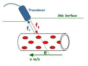
The Doppler effect is the change in the frequency of the ultrasound when it is reflected off a moving object e.g., red blood cells. If the blood cells are moving towards the transducer the frequency becomes higher, if moving away from the transducer the frequency is lowered.
Doppler measurements in medicine[edit | edit source]
During a Doppler sonography procedure, the Doppler effect can be observed in the reflection of ultrasound waves off reflecting structures in flowing blood such as erythrocytes. The frequency of reflected waves differs from the transmitted wave due to the movement of the reflector. If the blood flows towards the source of the waves - towards the ultrasonic probe, then the frequency of the reflected waves is higher than the transmitted frequency, if the blood flows in a direction away from the transducer, the frequency is lowered.
We do not measure the actual velocity but only the velocity component parallel to the probe. Therefore, if the blood flow measuring probe is located perpendicular to the vessel, it measures zero speed. Reflection occurs at the wall of the blood vessel and scattered from off the moving erythrocytes - the amount of waves that gets back to the probe is small (the blood is almost anechoic), but it is enough to determine the frequency shift; and from that it is possible to derive the flow rate of the blood as well as the nature of the flow (laminar, turbulent).
In general, Doppler measurements can be performed in two modes continuous wave (CW) or pulsed wave (PW). Small hand held Doppler devices such as the one used in this experiment use CW Doppler whilst 2D/3D Doppler imaging devices use PW systems. In PW systems the transmitter transmits in pulses. This mode allows you to not only measure the frequency change between the transmitted and received signals, but also the time the reflected signal takes to return to the probe. The time lag between the impulse sending and capturing its reflection, determines the depth at which the flow rate has been measured. The result is displayed as a two-dimensional image of measured speeds.
Blood flow parameters[edit | edit source]
Some blood flow parameters that can be measured are:
Maximum systolic velocity (S)- maximum velocity in systole.
Minimum diastolic velocity (D)- minimum velocity of the flow at the end of the diastole
Mean velocity V-mean.
S/D ratio (systolic / diastolic ratio)
Resistive index RI (resistance index) = (S-D)/S
Increase in RI indicates increased resistance to blood flow leading to a reduction of the diastolic velocity. The resistance index may provide information on the efficiency of the patients blood flow and whether arteries show abnormal properties or not . A value of ≈ 0.60 is considered as normal, however a value above 0.70 indicates that the patient may have a condition such as renal artery disease.
Pulse index PI (pulsatility index) = (S-D)/VMean
Different arteries have different values and factors such as age, fitness and even, as suggested by some studies, the emotional stage of the patient.
Heart rate HR (heart rate) is the number of heart beats per unit time, most often per minute. HR = 1 / T where T is the length of the heart period.
Protocol[edit | edit source]
During this practical, you will be working with a simple CW Doppler device used to measure the velocity of blood in arteries at a given location - the Bidop Hadeco (ES-100V3) portable Doppler ultrasonic measuring device. You can watch a video of the use of such a device here https://www.youtube.com/watch?v=Zq20f0z65Xg
If it is not already connected, connect the Bidop device to your computer using a USB cable. There are only two ports on the device with a directional label. To connect your device to the computer, use the port with the output symbol.
Run the Smart-V-Link software using the desktop icon. Fill out the data on the home screen. Disregard the automatically computed data, fill in all information manually (basic information is sufficient), and save. Continue to the program home page.
Remove the black cap from the probe.
Hold the small button on the probe to turn it on.
In Settings, set the device to communicate with the computer (COM). The access can be connected via any USB port on the computer. (In order for this option to be available, it is necessary to switch on the measuring device.) If the option does not appear even when the device is turned on, press Search Comm. Once found, return to the main screen.
Apply a thin layer of gel to the probe (approx. 3 mm thick).
For the purpose of this practical, the Individual Waveform is the most appropriate measurement setting. However, in practice there are alternative methods for data collection. (For example, with Venous Doppler it is possible to measure and compare venous flow in both limbs)
Double-click on the gray field on the screen and device will start to measure . The values will appear on the screen as a moving curve.
Place the probe at an angle of approximately 45° against the flow of blood and try to find the brachial pulse by the brachial artery situated near the elbow on the upper arm.
Turn the dial on the device to adjust the volume so that you can orient yourselves aurally as well. When you find the location of an artery, the probe will emit a whistle-like sound.
Hold the probe steadily in place and wait until the field on the screen is fully filled with a periodic waveform. Continue measuring until you have at least peaks, then end the measurement by briefly pressing the small button on the probe.
Press Decision. You will see the values at the top of the screen that you will later use to fill in your log. Save the graph via PrintScreen on the keyboard. This file can be printed directly.
Be sure to clean off any residual gel from the probe before leaving your station.
Processing of Individual Measurement Results
Once you have finished the measurements with Smart-V-Link, open the print screen by pressing the PrtSc key.
Import the PrintScreen graph into GIMP (Ctrl+v). From here, you will transfer your values into the Excel sheet.
Cut the image either by selecting the Trim tool (Ctrl+Shift+C) directly on the keyboard or by the path: Tools → Transformation Tools → Trimming and Dimensions.
Copy the “Doppler new” Excel spreadsheet to the desktop and rename it using the name of your group. Then, open it and write the measurement results from the in the yellow fields.
First calibrate. Place the cursor at point 0, where the pixel coordinates {X:Y} will appear in the lower left bar. Copy these coordinates to the first table. In a similar fashion, fill in all subsequent calibration values. (Note: the end of diastole is equal to the onset of systole.)
At the bottom right of the Excel sheet will be the manually retrieved values, which are calculated from the table macros. Fill in the yellow boxes next to the manual values with the automatic values recorded in the Smart-V-Link program. You can find these values at the top of your graph when you clicked the Decision tab.
Compare the results.
Save the file (File->Save as) in .bmp format on the desktop (for easier access). This makes the file ready for printing.
Print both the graph and the Excel table. Drag all saved files into the desktop folder labeled: “Results.”
Information for Automatic and Manually Measured Values
Measured Pulse Wave Parameters:
HR (Heart Rate)
RI (Resistance Index)
SD (Systolic / Diastolic ratio = S / D ratio)
MEAN (Mean Flow = average velocity [Vmean])
PI (Pulsatility Index = pulse index)
Important Notes for the Protocol[edit | edit source]
DO NOT use this device on the chest, abdomen, head, or neck area.
To prevent any unwanted reflection of ultrasound waves, apply a layer of gel to the contact tip of the probe about 3 mm thick. For the waves to penetrate better in a solid medium than air medium, the ultrasound gel enables the waves to transmit directly to the targeted tissue providing coupling between the skin and the probe as a type of conductive medium. Once the gel begins to dry, apply a new layer. However, if the gel covers too much dermal surface area, the probe may not record properly.
While using the probe, carefully and patiently locate the signal. The place for examination is good in advance, for example, as a place where you can feel the pulse with the fingers.
Do not move the probe after finding the appropriate probe location (unlike 2D/3D sonography).
The program may unexpectedly stop (program not responding). Typically, you can resolve the issue by restarting the program without further problems.
Protocol Preparation[edit | edit source]
To prepare for the protocol, use the form found under P8 - Doppler Practical on Moodle. Just before the start of taking measurements, fill in the boxes noting the order of the examiner and patient, the team number, the date, the start time of the examination, the investigated artery, and the investigated side (i.e.- sin means left, dx right). At the end of the experiment, record the end-time of the examination.
Write down the measured values from the experiment straight into the data table.
In the discussion, compare your calculated values with the automatically calculated and any theoretical assumptions you had about your measurements. Also, mention the fact that affects the outcome of the examination.
In the conclusion, briefly state whether the result of your measurement corresponds to a normal finding and whether a low-resolution or high-resolution curve was measured.
Conclusion[edit | edit source]
Over the course of the last half decade the development on the field of diagnostic ultrasound has been rapid, the newest technology being 3D-imaging. Doppler sonography improves diagnosis due to provision of immediate clinical information, hence the reduction of wrong diagnosis consequently a reduction of harm to the patient which could be caused by wrong treatments. On the financial side the immediate result reduce overall healthcare costs. All in all Doppler sonography is a great tool to use to provide initial provisional diagnoses, and a tool to give direction to further tests.
References[edit | edit source]
Kurt Hecher; “Doppler In Obstetrics”, 12 Dec 2018, https://fetalmedicine.org/var/uploads/Doppler-in-Obstetrics.pdf
Oriana Tolo; “The Doppler effect in medical ultrasound”, 12 Dec 2018 http://www.ultrasoundtraining.com.au/sb_cache/events/id/35/f
Brig. RS Moorthy; “Doppler Ultrasound- Editorial”, 12 Dec 2018, http://medind.nic.in/maa/t02/i1/maat02i1p1g.pdf
Wiley-Blackwell; “Doppler ultrasound in pregnancy reduces risk in high-risk group”, https://www.sciencedaily.com/releases/2010/01/100119213045.htm
Radiological Society of North America; “General Ultrasound” http://www.radiologyinfo.org/en/info.cfm?pg=genus
Jan G Aaranoudse; “Calculation of PI from a uterine artery Doppler flow velocity waveform“, 14 Dec 2018, https://www.researchgate.net/figure/Calculation-of-PI-from-a-uterine-artery-Doppler-flow-velocity-waveform_fig1_8992772
Duke Medical School; Anatomy Learning Recources”, 14 Dec 2018 https://web.duke.edu/anatomy/Lab12/Lab13_preLab.html


