Doppler sonography/medical applications
Introduction[edit | edit source]
Doppler sonography is the practice of using reflected waves to see how blood flows through a blood vessel. It helps doctors measure the velocity of blood through major, arteries and veins, such as those of the arms, legs, and neck.
The Doppler sonography is a non-invasive test. It estimates the blood flow of by bouncing high frequency sound waves off circulating red blood cells. A regular ultrasound (called B mode ultrasound) uses high frequency sound waves to produce images of anatomical regions of the body but does not measure blood flow.
A Doppler sonography can estimate how fast blood flows by measuring the change of frequency of the reflected ultrasound. The Doppler sonography is a preferred method to more invasive procedures such as arteriography and venography, which involve injecting dye into blood vessels, so they show up clearly on x ray images. These are less safe as they make use of ionizing radiation and moreover do not measure velocities.

Importance in Clinical Medicine[edit | edit source]
Doppler ultrasound is done to:
- find blood clots and blocked or narrowed vessels in almost any part of the body, especially in the neck, arms and legs. (Used in diagnosis of diseases such as venous insufficiency and atherosclerosis)
- Evaluate leg pain that may be caused by intermittent claudication, a condition caused by atherosclerosis of lower extremities.
- Evaluate blood flow after a stroke or other condition that might be caused by a problem with blood flow. Evaluate of a stroke can be done through a technique called trans cranial Doppler (TCD) ultrasound.
- Evaluate varicose veins or other vein problems.
- Map veins that may be used for blood vessel grafts. It can also check the condition of grafts used to bypass blockage in an arm or leg.
- Find out the amount of blood flow to a transplanted kidney or liver.
- Monitor the flow of blood following blood vessel surgery.
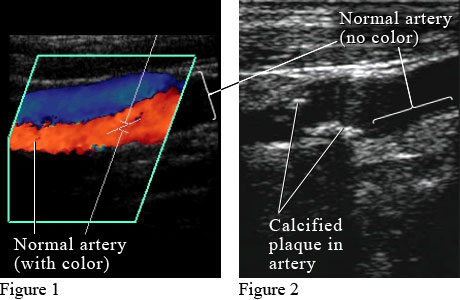
- Find out the presence, amount and location of arterial plaque. Plaque in the carotid arteries can reduce blood flow to the brain and may increase the risk of stroke.
- Guide treatment such as laser or radiofrequency ablation of abnormal veins.
- It can also reveal blood clots in leg veins (deep vein thrombosis or DVT) that could break loose and block blood flow to the lungs (pulmonary embolism).
- Check the health of a fetus. Blood flows in the umbilical cord, through the placenta, or in the heart and brain of the fetus may be checked. This test can show if the fetus is getting enough oxygen and nutrients. Doppler ultrasound may be used to guide decisions during pregnancy when:
- The fetus is smaller than normal for its gestational age. Blood flow through the large blood vessel in the umbilical cord (the umbilical artery) can be looked at.
- Rh sensitization has occurred. Blood flow through a blood vessel in the brain (the middle cerebral artery or MCA) can be used to monitor fetal health.
- The mother had problems, such as preeclampsia or sickle cell disease.
Different Types[edit | edit source]
- Bedside or continuous wave Doppler monitor: this type of Doppler sonography uses the change in frequency of the sound waves to provide information about the blood flow through a vessel. The doctor listens to the sounds produced by the transducer to evaluate the blood flow through an area that may be blocked or narrow
- Color Doppler (also known as Duplex Doppler): the velocities in the different patient voxels are visualized as a coloured image superimposed on the normal anatomical greyscale B-mode image. Different shades of red and blue are the most widely recognized hues to indicate the different velocities. The colours red and blue indicate the direction of the velocity (whether towards or away from the transducer).
- Power Doppler: Power Doppler can get some images that are hard or impossible to get using standard color Doppler. Power Doppler is used to indicate simply the presence or otherwise of blood flow. It does not give accurate velocities, however it detects very low blood flows.
Limitations of Doppler Sonography[edit | edit source]
- Vessels deep in the body are harder to see than superficial vessels. Specialized equipment or other tests such as CT or MRI may be necessary to properly visualize them.
- Smaller vessels are more difficult to image and evaluate than larger vessels
- Calcifications that may be present in vessels due to atherosclerosis may obstruct the ultrasound beam.
- Sometimes ultrasound cannot differentiate between a blood vessel that is completely closed of and one that is narrowed. Even if there is a small opening, the weak blood flow sometimes produces an undetectable signal.
Conclusion[edit | edit source]
The eventual fate of Doppler sonography is solid. The Doppler ultrasound method has been developed and extensively used in the medical field, especially in cardiology, where it is necessary to measure time dependent velocity profiles inside a blood vessel in order to determine the flow rate and character of blood flow. The expansion of miniaturized scale air pocket contrast ogents which are injected into the circulation system, improves the returning Doppler signs, which makes vastly improved pictures of stream in the heart and little veins.
Doppler is also helping in the delivery of chemotherapy. The administrator watches with Doppler for the contrast agent bubble's landing in the tumour, and then blasts them inside of the tumour. Another new therapeutic use of Doppler ultrasound is thrombolysis of blood clots in the brain. While thrombolysis does not rely upon the Doppler effect, colour Doppler is used to guide the application of the ultrasonic energy to specific blood vessels within the brain. The sonic vibrations appear to enhance the thrombolytic actions of anticoagulants. The first 50 years of the application of ultrasound through sonography and Doppler imaging has been an amazing period in modern medicine. As the population ages, there will only be an expansion of these techniques to improve the health of all people from before birth to old age.
Links[edit | edit source]
References[edit | edit source]
- Walker, Andrew et al. ACCURACY OF SPECTRAL DOPPLER FLOW AND TISSUE VELOCITY MEASUREMENTS IN ULTRASOUND SYSTEMS. 1st ed. Linköping, Sweden: N.p., 2015. Print.
- (ACR), Radiological. "Venous Ultrasound - Extremities". Radiologyinfo.org. N.p., 2015. Web. 18 Dec. 2015.
- WebMD,. "Doppler Ultrasound". N.p., 2015. Web. 18 Dec. 2015.


