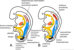
Development of the urinary system
The development of the urinary and sexual systems is closely related. They develop together from the intermediate mesoderm (it runs along the back wall of the abdomen) and their ducts initially open into a common cavity - the 'cloaca.
Urinary System[edit | edit source]
During prenatal human development, 3 partially overlapping systems of excretory organs are established:
- Pronephros - is a rudimentary and non-functional organ.
- Mesonephros - is functionally applied only in a short period of time, in the early fetal period.
- Metanephros - which is the definitive kidney.
Pronephros[edit | edit source]
Pronephros also foreskin develops during 4. weekly. This organ is visible at the beginning of the fourth week as 7-10 distinct groups of cells in the neck region. These clusters of cells are considered rudimentary excretory units – 'nephrotomes. These structures are no longer visible by the end of the 4th week, with the cranially lying nephrotomes disappearing before the caudally lying ones are formed.
Mesonephros[edit | edit source]
The mesonephros, also known as the primary kidney, consists of a group of excretory ducts and the ductus mesonephricus, which originate "from the intermediate mesoderm at the level from the upper thoracic to the upper lumbar region" (L3 segment). The first excretory ducts appear at the time of the disappearance of the pronephros – at the beginning of the 4th week of development. These ducts rapidly elongate, acquire an S-shaped loop, and contact a ball of capillaries that form a glomerulus' at their medial end. The excretory ducts form around the glomerulus Bowmann's capsule and together with it form a structure called the corpusculum renale. Laterally, the excretory ducts open into a longitudinal draining duct – 'ductus mesonephricus', also called 'ductus Wolffi'. The mesonephros is a large ovoid paired organ on the sides of the midline in the middle of the second month. Here it lies lateral to the developing gonad, and the mound formed by both these organs forms the fold, the 'plica urogenitalis'. Gradually, the canals and glomeruli of the mesonephros disappear in the craniocaudal direction, while at the end of the 2nd month of development, most of the mesonephros is already degenerated in this way. Only in male fetuses several caudal ducts and ductus mesonephricus persist so that they can participate in the formation of the excretory gonads, this does not happen in female fetuses.
Metanephros[edit | edit source]
The final excretory organ appears in the 5th week. Its excretory units develop in the same way as the mesonephros, from the metanephrogenic blastema - an unsegmented mass of intermediate mesoderm tissue in the lower thoracic, lumbar, and sacral regions. However, the development of the system of collection and drainage channels is different. The collecting and efferent ducts of the definitive kidney are formed 'by the end of the 5th month of development, developing 'from the ureteric bud' which arises from the mesonephric duct near its mouth into the cloaca. The bud grows into the metanephros tissue, which forms a cap above it. As the bud subsequently expands, it forms a primitive 'renal pelvis' and divides into a cranial and caudal part, the basis of the next 'calices majores'. Each calyx further forms 2 new buds that penetrate the tissue of the metanephros and continue dividing until a min. 12 generations of channels. However, the appearance of the canals created in this way changes, as the canals of the 2nd order enlarge and pull the canals of the 3rd and 4th generation into each other, creating 'calices minores'. Furthermore, the collecting and draining ducts of the 5th and higher generations lengthen considerably and at the same time converge to the calices minores to form pyramides renales'. The ureteral bud thus gives rise to the ureter, renal pelvis, small and large calyces, as well as collecting and draining ducts, of which 1–3 million are established.
Excretory System[edit | edit source]
At the periphery of each newly formed collecting duct we find a ``metanephros tissue cap. The collecting ducts induce the formation of small vesicles in this tissue – vesicules renales, which are formed by metanephros cells. From the vesicules renales, ``channels twisted into the shape of the letter S gradually arise, then capillaries enlarge around the ace-shaped channel and form balls - 'glomeruli'. These channels and their glomeruli are the basis of the excretory unit - the nephron. From the peripheral end of the nephron, Bowman's capsule is formed around the glomerulus, and the glomerulus is eventually taken into it. The opposite end of the canal opens into the collecting duct, allowing passage from the Bowman's capsule into the drainage system. Subsequently, the excretory duct grows in length to form the proximal convoluted tubule, the loop of Henle and the distal convoluted tubule.
- The kidney therefore arises from 2 foundations
- from mesodermu metanephros – excretory unit;
- from the ureteral bud – collection and drainage system.
Nephrons are formed only in the prenatal period and there are approximately 1 million of them at birth. Urine begins to form from the 10th week of development not long after the capillaries of the glomeruli have begun to differentiate.
Molecular mechanisms of kidney development[edit | edit source]
Differentiation of the kidney is conditioned by the interaction of the epithelium (epithelium of the ureteric bud) and the mesenchyme (mesenchyme of the metanephrogenic blastema). Mesenchyme expresses the transcription factor WT 1, which is responsible for the ability of the metanephrogenic blastema to respond to the inductive influence of the ureteral bud. Mesenchyme also produces a number of other growth factors that mediate the interaction between epithelium and mesenchyme.
Bladder and urethra[edit | edit source]
During the 4th-7th week of development, the septum urorectale divides the cloaca into the sinus urogenitalis (front) and the canalis analis (back). The septum urorectale is a layer of mesoderm between the primitive anal canal and the sinus urogenitalis.
There are 3 parts to the urogenital sinus.
- The upper and largest part is the basis of the 'bladder'. The latter is initially related to the allantois canal, which represents the 'urachus'. After the lumen of the allantois is obliterated, a fibrous band is preserved from it from the top of the bladder to the navel - 'ligamentum umbilicale medianum'.
- Another part of the urogenital sinus is its "pelvic segment", from which the "prostatic and membranous part of the urethra" originates in males and the "whole urethra" in females. .
- The last part of the urogenital sinus is its "spongy part". From that in the male sex arises the spongy part of the urethra except for its pars glandis, which arises from the ectoderm epithelial plug. In female fetuses, this part gives rise to the vestibulum vaginae.
- During cloacal differentiation, the caudal sections of the ductus mesonephricus are drawn into the wall of the bladder so that the ureters now empty into the bladder separately. As the kidneys rise, their mouths move cranially, while the mouths of the mesonephric ducts move closer to each other, and in males they open into the prostatic part of the urethra as the ductus ejaculatorii. In females, the "ductus mesonephricus" "disappears" above the distance of the ureteral bud.
Urethra[edit | edit source]
The urethra epithelium originates in both sexes from the entoderm. The ligamentous and smooth muscle tissue that surrounds the urethra is 'derived from the mesoderm of the splanchnopleura'. Towards the end of the 3rd month the epithelium of the prostatic part of the urethra begins to proliferate to lay the foundations of the 'prostate in male fetuses and ' 'urethral and paraurethral glands in female fetuses.
Links[edit | edit source]
Related Articles[edit | edit source]
References[edit | edit source]
- SADLER, Thomas W. Langman's Medical Embryology. 1. edition. Grada, 2011. 432 pp. ISBN 978-80-247-2640-3.
- MOORE, Keith L – PERSAUD, T.V.N. The Birth of Man : Embryology with a clinical focus. 1. edition. 2000. 564 pp. ISBN 80-85866-94-3.

