Development of lymphatic vessels, nodes and spleen
The lymphatic system develops from the fifth week of intrauterine development (in contrast to the cardiovascular system, which begins its development already in the middle of the third week). There are two theories describing the development of lymphatic vessels. According to the first, centrifugal theory, lymphatic vessels are formed by budding of the endothelium of already existing veins . According to the second, centripetal, they arise by differentiation from mesenchyme in situ . Accepting both theories, it is probable that the deep lymphatic vessels are formed by budding and the superficial lymphatic vessel by differentiation from the mesenchyme.
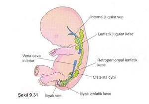
At the beginning of the development of the lymphatic system, 6 primary lymph sacs are formed:
- 2 jugular lymphatic bags - at the junction of the subclavian vein and the anterior cardinal vein,
- 2 iliac lymph sacs - at the junction of the iliac vein and the posterior cardinal vein,
- retroperitoneal lymph sac - on the back wall of the abdomen in the radix mesentery,
- cisterna chyli – dorsal to the retro peritoneal sac.
Development of lymphatic vessels[edit | edit source]
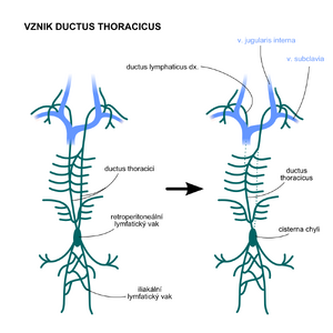
The emerging lymphatic vessels run along the veins and connect to the lymph sacs. The jugular sacs are connected by lymphatic vessels to the head, neck and upper limbs, the iliac sacs to the lower limbs, the retroperitoneal sac and the chyli cisterna to the primitive intestine. The jugular lymph sacs and the cisterna chyli are connected by two large channels – the right and left ductus thoracicus. These channels bring lymph to the bloodstream at the angulus venosus, which is the angle between the jugular vein and the subclavian vein. An anastomosis soon forms between the two canals, which runs craniocaudally from left to right. Later, the caudal part of the left thoracic d. and the cranial part of the right thoracic d. disappear and their function is taken over by a single ductus thoracicus. It is therefore formed by the caudal part of the right thoracic d., a common anastomosis and the cranial part of the left thoracic d. The ductus lymphaticus dexter arises from the upper part of the right d. thoracicus.
Development of lymph nodes[edit | edit source]
Lymph node development is divided into primary and secondary lymph node development. Lymphatic follicles are formed in them much later, almost before birth, and this is because their defensive function during intrauterine development is performed by the mother's immune system by creating antibodies that diffuse through the placenta.
- Primary lymph nodes – arise from the transformation of lymph sacs, which results in the greatest concentration of nodes precisely in the places of former lymph sacs. There is a penetration of mesenchymal cells into the lymphatic sacs, the cavities of which are subsequently transformed into a network of lymphatic channels. These ducts are the precursors of the future lymphatic sinuses inside the nodes. From other mesenchymal cells, the fibrous stroma and capsule of the nodules are subsequently formed. The only exception is the chyli cisterna, the upper part of which persists throughout life as a 50 x 6 mm sac.
- Secondary lymph nodes – arise along the lymphatic vessels. Thanks to this, there are lymph nodes in the course of each lymphatic vessel.
Development of the spleen[edit | edit source]
The development of the spleen is linked to the development of the gastrointestinal tract. The spleen arises from a cluster of mesenchymal cells in the dorsal mesentery of the stomach ( dorsal mesogastrium ) at the beginning of the 5th week. The spleen is first renculized, but before birth the renculization disappears and its remains are only visible as projections on its surface. Initially, the spleen also has a hematopoietic function, which is then taken over by the bone marrow.
The nascent spleen in the dorsal mesogastrium is displaced to the left due to the rotation of the stomach . This results in the attachment of the dorsal mesogastrium to the parietal peritoneum, and its remaining portion persists as the ligamentum lienorenale.
Developmental anomalies of the lymphatic system[edit | edit source]
- Diffuse swelling of parts of the body.
- There is a dilation of the primitive lymphatic channels and thus an accumulation of lymph in these areas of the body. Lymphedema can also be caused by hypoplasia of lymphatic vessels.
- Hygroma cysticum.
- These are large swellings in the inferolateral parts of the neck containing large cavities filled with fluid. They arise from the abnormal transformation of the jugular lymph sacs by their splitting off. These cavities are usually only noticeable in childhood.
Links[edit | edit source]
Related articles[edit | edit source]
- General anatomy of the lymphatic system • Lymph nodes • Spleen
- Spleen (preparation) • Node (preparation) • Thymus (preparation) • Tonsil (preparation)
References[edit | edit source]
- SADLER, Thomas W. Langmanova lékařská embryologie. Grada Publishing. 2011. 432 s. ISBN 978-80-247-2640-3.
- MOORE, Keith L. a T.V.N. PERSAUD. Zrození člověka : Embryologie s klinickým zaměřením. ISV nakladatelství. 2000. 564 s. ISBN 80-85866-94-3.
- ČIHÁK, Radomír. Anatomie 3. Druhé, upravené a doplněné vydání. Grada Publishing. 2004. ISBN 978-80-247-1132-4.


