Deformities of the lower limbs

Deformities of the lower limbs can be divided according to the place where they occur. They can be either congenital or acquired during life (overuse of the limb, trauma, metabolic disease, inflammation, etc.).
Deformities of the knee joint[edit | edit source]
Genua valga[edit | edit source]
Genua valga (bowed knees) are characterized by an increase in the physiological valgusness of the knee joints.
More detailed information can be found on the Genua valga page .
Genoa vara[edit | edit source]
Genua vara (bent knees) represent a very common deformity of childhood. They are normal in newborns. When the child begins to walk, the deformity becomes even more pronounced.
More detailed information can be found on the Genua vara page .
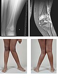
Genu recurvatum[edit | edit source]
A less common defect. It used to be associated with rickets, but today it is more likely to occur with disorders of the epiphyseal cartilage, such as inflammation, injury or after polio.
Clinically , hyperextension of the knee joint is present . The lower leg and thigh form an open angle forward.
Small defects do not need to be treated. If the stability of the limb is disturbed, we choose a surgical solution in the form of an osteotomy in the supracondylar region of the femur and also an operation of the surrounding soft tissues.
Genu flectum[edit | edit source]
It most often occurs in children with cerebral palsy . In adults, it is usually associated with deforming arthrosis of the knee joint, which is associated with the formation of a varus position of the knee, or with rheumatoid arthritis , which is associated with vlagous involvement and flexion contracture.
Clinically, we see the knee in flexion , while full extension cannot be achieved.
Therapy depends on the cause of the disease. In case of cerebral palsy, we choose the prolongation of the flexor tendons of the knee joint. Total joint replacement is required in adults with deforming arthrosis and flexion contracture .
Genua valga 
Genoa vara

Genu recurvatum


Leg deformities[edit | edit source]
Equinus dog[edit | edit source]
Czech foot vertical . It is a deformity that arises from disorders of the nervous system in spastic paralysis, weak paralysis after poliomyelitis or paralysis of the extensors of the leg.
Clinically, the foot is usually fixed in plantar flexion . The patient steps on the front part of the foot, is unable to step on the heel. Such walking leads to the collapse of the transverse arch and the formation of pressure marks on the tread surface. There is atrophy of the calf and leg muscles.
Treatment can be conservative, which includes rehabilitation, exercises for the shortened Achilles tendon, or the use of a corrective cast.
The Achilles tendon can be surgically lengthened.
Pes excavatus[edit | edit source]
Clubfoot usually has neurological causes (myelodysplasia, muscular dystrophy), Friedreich's disease, or arises secondary to inflammation .
Clinically, the longitudinal arch is accentuated. A contracture of the plantar fascia and soft tissues in the plantar area occurs. The fingers tend to have a claw-like position and bruises occur in the area of the metatarsal heads. The heels are varus.
Before therapy, it is necessary to send the patient for a neurological examination. It can be treated conservatively with special orthopedic shoes.
The surgical solution consists in releasing the plantar structures from the heel bone, or a wedge-shaped corrective osteotomy is performed.
Dog planovalgus[edit | edit source]
Longitudinally flat foot. There is an abnormal reduction or disappearance of the longitudinal arch. The heel is in a valgus position, the talus descends and the forefoot abducts.
More detailed information can be found on the Pes planovalgus page .
Dog transversoplanus[edit | edit source]
Transversely flat feet usually occur after the age of 30, and more often in women, who more often choose unsuitable shoes (short tight toes). In the wrong shoes, the forelegs are overloaded, which is further aggravated by possible excess weight .
Clinically, there is pain in the region of the heads of the metatarsals, sometimes also neuralgic pain from oppression of the plantaris medialis nerve (radiating between the III and IV toes). The pain usually subsides when the load is lightened. There is a gradual lowering of the arch due to weakening of the ligaments and muscles. The front part of the foot expands and the heads of the metatarsals arch into a flat surface, under which pressure marks are formed.
Transversely flat foot usually occurs together with flat foot, so therapy is usually linked to both of these deformities. Most often, we choose orthopedic insoles , or hearts for shoes. You can also use physical therapy , hydrotherapy and whirlpools. Sprays can be used for neuralgia .
If the pain does not subside, it is necessary to choose an operative solution, which consists in releasing the nerve by cutting the front edge of the intermetatarsal plantar ligament. For more severe defects (rheumatic involvement), we resect heads II.-V. metatarsus
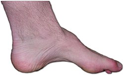
Pes excavatus 
Pes planovalgus


Deformities of the toes[edit | edit source]
Hallux valgus[edit | edit source]
A bunion is one of the most common foot deformities. It is much more common in adults as a result of weakening of the fibrous and muscular apparatus, which leads to a decrease in the transverse arch and a change in the position of the thumb.
More detailed information can be found on the Hallux valgus page .
Hallux varus[edit | edit source]
Sprained thumb. Unlike valgus, it is not as common.
See the Hallux varus page for more detailed information .
Hallux rigidus[edit | edit source]
Or a stiff thumb. It most often arises on the basis of arthrosis in the metatarsophalangeal joint of the thumb. It occurs in some professions or after inflammation and operations.
More detailed information can be found on the Hallux rigidus page .
Digitus malleus[edit | edit source]
This deformity usually arises in conjunction with hallux valgus and pes transversoplanus. We distinguish between two forms, the hammer finger and mallet finger.
See the Digitus malleus page for more detailed information .

Hallux valgus 
Hallux varus 
Hallux rigidus


Disease in the area of the heel[edit | edit source]
Haglund's exostosis[edit | edit source]
Exostosis arising on the back of the heel bone due to long-term mechanical irritation.
See Haglund's Exostosis for more detailed information .
Calcar calcanei[edit | edit source]
In other words, a spur is a deformity in the area of the heel bone. It most often occurs between the ages of 40 and 60 and is present bilaterally.
See Calcar calcanei for more detailed information .

Calcar calcanei 
Haglundova exostosis

.jpg)
Links[edit | edit source]
Reference[edit | edit source]
References[edit | edit source]
- SOSNA, A., P. VAVŘÍK and M. KRBEC, et al. Basics of orthopedics. 1st edition. Prague: Triton, 2001. ISBN 80-7254-202-8 .