Congenital defects of the external and middle ear

Anomalies of the external and middle ear are related to the maldevelopment of the first gill slit and the first two gill arches. These diseases are manifested by shape deformities of the auricle and ear canal. They can occur together with dysostoses of the lower jaw, zygomatic bone and auditory ossicles.
Congenital malformations of the auricles relate to shape, size and position.
Otapostasis[edit | edit source]
The most common congenital defect is otapostasis, the so-called standing bolt. It is an inherited, frequent defect of the auricle, the essence of which is the smoothing of the fold of the antihelix. The defect can be corrected surgically (the most suitable time for correction is between 5 and 6 years). The principle is the creation of a new antihelix.
Microtia[edit | edit source]
Another very common defect is microtia, i.e. reduction of the auricle. The defect can be classified into 3 degrees, in the case of the 2nd and 3rd degrees, the bolt is not only reduced in size, but also deformed in shape.
Anotia[edit | edit source]
Anotia is the complete failure of the auricle to develop.
Preauricular appendices[edit | edit source]
Preauricular appendices are usually unilateral appendages of the pinna. They mostly have the shape of bumps or pedunculated lobes located in front of the tragus.
Congenital ear fistula[edit | edit source]
These are canals mainly in the ascending part of the helix, external fistulas are less common, on the neck they are colo-auricular fistulas. Their danger lies in possible secondary infection, so they need to be surgically removed.
Stenosis and atresia of the auditory canal[edit | edit source]
Stenosis is a narrowing of the ear canal. Atresia is a complete closure of the ear canal - the ear canal is completely impassable. Atresia can be both bony and fibrous. It is the cause of conductive hearing loss. Both defects (stenosis and atresia) can be associated with other defects (auricle disorder, middle ear disorder, facial nerve disorder ). The therapy is surgical. It consists in the canalization of the ear canal, or the reconstruction of the middle ear.

Otapostasis

Antihelix after reconstruction
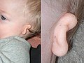
Microtia

Atresia


Links[edit | edit source]
Source[edit | edit source]
- BENEŠ, Jiří. Study materials [online]. ©2007. [cit. 2009]. <http://jirben2.chytrak.cz/materialy/orl_jb.doc>.
References[edit | edit source]
- KLOZAR, Jan, et al. Speciální otorinolaryngologie. 1. edition. Prague : Galén, 2005. 224 pp. ISBN 80-7262-346-X.
Related Articles[edit | edit source]
| This article is a stub. You can join the authors and edit it. You can discuss the changes at discussion. |