Colles fracture
From WikiLectures

This article has been translated from WikiSkripta; ready for the editor's review.
A Colles fracture is caused by a fall on a dorsiflexed and pronated arm :
- the radius breaks 2–3 cm proximal to the carpal joint ,
- the distal fragment dislocates dorsally and radially.
In half of the cases there is also a fracture of the ulna styloid process. Age-wise, it occurs in two peaks:
- at a younger age is related to increased activity,
- in old age, it is related to osteoporosis (along with femoral neck fractures and vertebral compression fractures).
Content[edit | edit source]
- 1Clinical picture and diagnosis
- 2Therapy
- 3Complication
- 4Links
- 4.1related articles
- 4.2Source
Clinical picture and diagnosis[edit | edit source]
Colles fracture.
- typical bayonet-like position when viewed from above, fork-like position when viewed from the side,
- pain, swelling, disfigurement of the wrist, limited mobility in the wrist,
- on the X-ray, we assess the inclination of the articular surface of the radius (30° in the antero-posterior projection, 15° in the lateral view – it decreases in the case of a fracture),
- may be:
- fracture of the processus styloideus radii ,
- rupture of the ulnar collateral ligament ,
- luxation of the radio-ulnar joint ,
- the fracture can also be comminuted (shattering) .
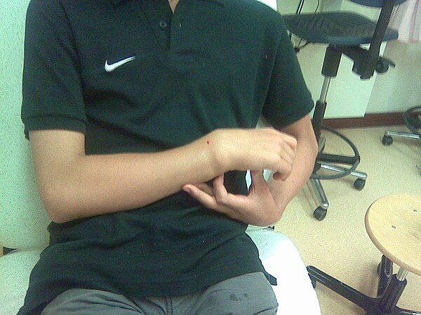
Position of the wrist joint.
Therapy[edit | edit source]
- Conservative (most are treated conservatively)
- local anesthesia (10 ml of 1% mesocaine to the hematoma site),
- reposition – pull for the thumb in the axis of the joint, for the other fingers in the direction of ulnar duction with a flexed elbow for a counter-pull (finger cups are suitable
- apply a dorsal plaster cast from the elbow to the heads of the metacarpals in slight wrist flexion and ulnar duction,
- should follow :
- x-ray check,
- finger blood flow control,
- in 2 days check to finish the cast (with X-ray),
- another X-ray check after 1 week and after 3 weeks,
- immobilization 6 weeks – immobilization in ulnar duction and palmar flexion,
- inadequate position after reduction:
- shortening of the radius by more than 2 mm,
- dorsal angulation above 5°,
- volar angulation above 20°,
- deficit on the articular surface of the radius above 1 mm.
- Operating :
- in these cases :
- if repositioning fails ,
- intra-articular fractures ,
- open fractures,
- options are:
- percutaneous fixation with Kirschner wires during closed reduction,
- external fixation,
- mini-incision tension screws,
- open reposition with a T-plate,
- LCP (locking compression plate).
- After surgery to stabilize the joint with an orthosis, full recovery in 10 weeks .
- in these cases :
In elderly people with osteoporosis, it is sometimes better not to attempt a reduction due to further possible disruption.
Complication[edit | edit source]
- shape changes in the wrist due to secondary redislocation and permanent difficulties in joint movement, which sometimes need to be solved by osteotomy and shortening of the ulna;
- rupture of the extensor pollicis longus tendon;
- carpal tunnel syndrome.
Links[edit | edit source]
[edit | edit source]
- Fractures of the forearm
- Radius
- Ulna
Source[edit | edit source]
- PASTOR, Jan. Langenbeck's medical web page [online]. [feeling. 2009]. < https://langenbeck.webs.com/ >.
- ZEMAN, Miroslav, et al. Special surgery. 2nd edition. Prague: Galén, 2006. 575 pp. ISBN 80-7262-260-9.