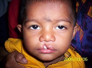
Cleft defect
Cleft defects (schisis) are the most common Congenital developmental defects. According to localization, we distinguish between: cheiloschisis (cleft lip), palatoschisis (cleft palate), gnathoschisis (cleft jaw), combined cheilognathoschisis (cleft lip and jaw) and total cleft cheilognathopalatoschisis (cleft lip, palate and jaw).
In addition to cleft lip, palate, and jaw, there are other types of clefts such as spinal clefts and abdominal wall clefts.
Etiology and pathogenesis[edit | edit source]
The annual incidence of cleft defects in newborns is around 1.8 per 1000 births. A general cleft is more common in boys, isolated more often in girls. The left side of the face is damaged twice as often as the right. In European and Mongoloid races, the occurrence is more frequent than in African Americans.
They arise roughly in the 6th–9th century. week of pregnancy, when the central and lateral parts of the face do not connect. The most critical is the 8th week of pregnancy, when the fusion of the medial nasal processes with the maxillary processes (and the horizontalization of the palatal plates of the secondary palate) occurs. The range can be different, from a hint to a bilateral defect, accompanied by difficulty in taking food and a speech disorder.
It is a multifactorial disease where heredity plays an important role, but medical conditions of the mother, such asdiabetes or epilepsy are also a major factor.
A preventive measure is an increased intake of folic acid especially in the first trimester. It is also necessary to limit exposure to teratogens (alcohol, medications, nicotine, drugs, infectious diseases, radioactive radiation, etc.).
Types of cleft defects[edit | edit source]
We distinguish defects in the face, neural tube and abdominal wall
Facial clefts[edit | edit source]
- Cleft lip
- Only the lip is affected, the jaw and palate are fine. It looks like a notch in the upper lip and can extend up to the nose. Clefts can be unilateral or bilateral, rarely clefts of the lower lip appear.
- Can be easily corrected surgically, operations during the first days of a newborn's life usually do not leave scars.
- Cleft palate
- In a cleft palate, the two bony parts of the palate do not fuse together. Cleft of the soft palate and other parts of the oral cavity rarely occurs. An opening in the upper part of the oral cavity, can be seen in the mouth of the affected child , which can make breathing and breastfeeding difficult.
- Isolated cleft palate
This type of cleft occurs less often than classic cleft lip and is more common in girls (in 67%), the incidence of this defect does not increase with the age of the mother. In girls, both palatal processes join a week later (a more frequent occurrence) than in boys.
- It is known that certain drugs, such as antiepileptic drugs , given in early pregnancy increase the risk of cleft palate.
Spina bifida[edit | edit source]
Developmental anomaly of the spine and spinal cord. In a cleft, neural tube defects occur so that the vertebrae of the spine do not close into an arch. A slit is created through which the spinal cord or nerves can arch. This type of cleft is less common and occurs in about 1-2 babies out of 1000 pregnancies. According to damage to the vertebrae, we distinguish 3 types: spina bifida occulta, meningocele, meningomyelocele.
- Spina bifida occulta
- Occult spina bifida, where the spinal arch is split and the spinal cord is intact. This type of cleft is common and is usually discovered incidentally through an X-ray examination . Symptoms include darker skin color and significant hair growth oever the affected area
- Meningocele
- The spinal cord is pushed through the opening in the vertebrae and the spinal cord is not broken. Individuals with this type may have mild disabilities.
- Meningomyelocele
- The most serious type of spina bifida. The spinal cord is pushed out as well as the spinal cord through the opening in the vertebrae. The spinal cord and its sheaths are covered by skin, the spinal cord together with the nerves may be exposed. In this type, the spinal cord is always damaged and this leads to paralysis of the lower limbs and the inability to control the bladder and anal sphincter.
- Children mostly die from meningitis or urinary tract infections.
Abdominal wall cleft[edit | edit source]
This type of cleft defect occurs in the first trimester of pregnancy. "Intestinal loops" migrate temporarily outside the abdominal cavity towards the attachment of the umbilical cord in each fetus. Under normal circumstances, it returns to the abdominal cavity around the tenth week of pregnancy. In this defect, it is typical that there is a defect in the return of intestinal loops and the abdominal wall does not close. Most cases of abdominal wall clefts are sporadic and cannot be directly affected.
Complications of cleft defects[edit | edit source]
A common complication of cleft defects is inflammation of the middle ear cavity . The reason may be the involvement of the Eustachian tube , which, if damaged, spreads bacteria and causes inflammation. Another complication can be food intake disorders, where milk and other food flows into the nasal cavity.
Links[edit | edit source]
Related Articles[edit | edit source]
References[edit | edit source]
- SADLER, T.W. Langman's Medical Embryology. 10th edition. vydavatel, 2006. 385 pp. ISBN 978-0-7817-9485-5.
- VACEK, Zdeněk. Embryologie. 1st edition. 2006. 256 pp. ISBN 978-80-247-1267-3.
- MUNTAU, Ania. Pediatrie. 2nd edition. Grada, 2014. ISBN 978-80-247-4588-6.

