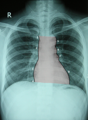
Cardiac silhouette
The heart is displayed on the X-rays in anterior (standing), anteroposterior (lying down) and oblique projections. Various dimensions can be read from the image of the cardiac silhouette, such as the maximum distance from the midline (left and right), as well as the radiological length of the heart, which is the distance from the border of superior vena cava and the right atrium to the left ventricle and diaphragm. According to the deviation from the horizontal, we measure the inclination of the heart, which is most often 45 °.
Front projection[edit | edit source]
The following are recognizable on the heart shadow:
- short part of v.brachiocephalica dextra,
- v. cava superior,
- to the diaphragm reaching atrium dextrum,
- (only with deep inspiration v. cava inferior is visible),
- arcus aortae,
- truncus pulmonalis (along with a. pulmonalis sinistra),
- auricula sinistra,
- ventriculus sinister, reaching to the diaphragm into the medioclavicular line.
Oblique projections[edit | edit source]
The Right oblique projection is also called fencing. It mainly shows the right part of the heart. The space behind the cardiac silhouette (Holzknecht's field) is also evaluated.
The left so-called boxing projection shows both ventricles, the aortic arch and the pulmonary artery.
Links[edit | edit source]
Related links[edit | edit source]
Bibliography[edit | edit source]
- ČIHÁK, Radomír – GRIM, Miloš. Anatomie 3. 2., upr. a dopl edition. Praha : Grada, 2004. 673 pp. ISBN 80-247-1132-X.

